Pedro Barata, Mariana Cervaens, Rita Resende, O ́scar Camacho and Frankim Marques
Abstract: In the last decade, competitive sports have taken on a whole new meaning, where intensity has increased together with the incidence of injuries to the athletes. Therefore, there is a strong need to develop better and faster treatments that allow the injured athlete to return to competition faster than with the normal course of rehabilitation, with a low risk of re-injury. Hyperbaric therapies are methods used to treat diseases or injuries using pressures higher than local atmospheric pressure inside a hyperbaric chamber. Within hyperbaric therapies, hyperbaric oxygen therapy (HBO) is the administration of pure oxygen (100%) at pressures greater than atmospheric pressure, i.e. more than 1 atmosphere absolute (ATA), for therapeutic reasons. The application of HBO for the treatment of sports injuries has recently been suggested in the scientific literature as a modality of therapy either as a primary or an adjunct treatment. Although results have proven to be promising in terms of using HBO as a treatment modality in sports-related injuries, these studies have been limited due to the small sample size, lack of blinding and randomization problems. HBO seems to be promising in the recovery of injuries for high-performance athletes; however, there is a need for larger samples, randomized, controlled, double-blinded clinical trials combined with studies using animal models so that its effects and mechanisms can be identified to confirm that it is a safe and effective therapy for the treatment of sports injuries.
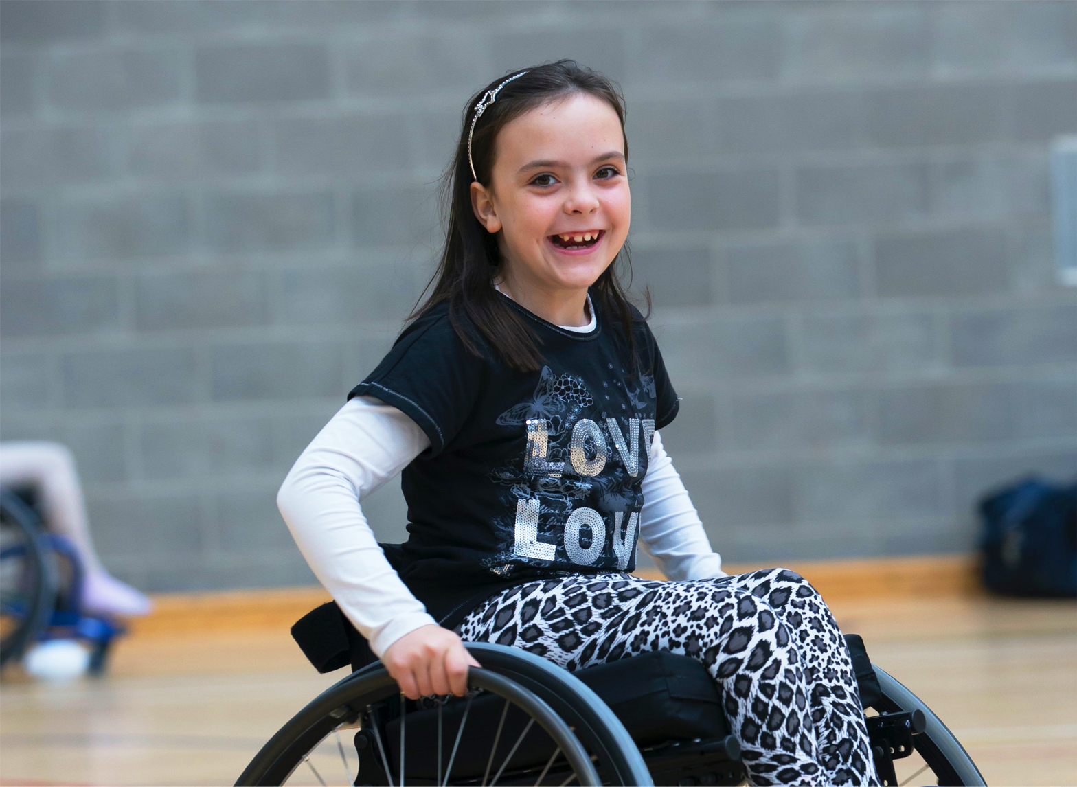
In the last decade, competitive sports have taken on a whole new meaning, where intensity has increased together with the incidence of injuries to the athletes. These sport injuries, ranging from broken bones to disrupted muscles, tendons and ligaments, may be a result of acute impact forces
in contact sports or the everyday rigors of training and conditioning [Babul et al. 2003].
Therefore, a need has emerged to discover the best and fastest treatments that will allow the injured athlete to return to competition faster than the normal course of rehabilitation, with a low risk of re-injury.
Hyperbaric oxygen therapy (HBO) is the therapeutic administration of 100% oxygen at pressures higher than 1 absolute atmosphere (ATA). It is administered by placing the patient in a multiplace or in a monoplace (one man) chamber and typically the vessels are pressurized to 1.53.0 ATA for periods between 60 and 120 minutes once or twice a day [Bennett et al.2005a]. In the monoplace chamber the patient breathes the oxygen directly from the chamber but in the multiplace chamber this is done through a mask. At 2.0 ATA, the blood oxygen content is increased 2.5% and sufficient oxygen becomes dissolved in plasma to meet tissue needs in the absence of haemoglobin-bound oxygen, increasing tissue oxygen tensions 10-fold (1000%) [Staples and Clement, 1996]. HBO is remarkably free of untoward side effects. Complications such as oxygen toxicity, middle ear barotrauma and confinement anxiety are well controlled with appropriate pre-exposure
orientations [Mekjavic et al. 2000].
HBO has been used empirically in the past, but today information exists for its rational application. This review aims to analyse the contribution of HBO in the rehabilitation of the different sports injuries.
Hyperbaric Oxygen Therapy
Hyperbaric therapies are methods used to treatdiseases or injuries using pressures higher than local atmospheric pressure inside a hyperbaric chamber. Within hyperbaric therapies, HBO is the administration of pure oxygen (100%) at pressures greater than atmospheric pressure, i.e. more than 1 ATA, for therapeutic reasons [Albuquerque e Sousa, 2007].
In order to be able to perform HBO, special facilities are required, with the capacity for withstanding pressures higher than 1 ATA, known as hyperbaric chambers, where patients breathe 100% oxygen [Fernandes, 2009]. In the case of single monoplace chambers (with a capacity for only one person) the oxygen is inhaled directly from the chambers’ environment [Fernandes, 2009]. Although much less expensive to install and support, they have the major disadvantage of not being possible to access the patient during treatment. It is possible to monitor blood pressure, arterial waveform and electrocar diogram noninvasively, and to provide intravenous medications and fluids. Mechanical ventilation is possible if chambers are equipped appropriately, although it is not possible to suc- tion patients during treatment. Mechanical ventilation in the monoplace chamber is provided by a modified pressure-cycled ventilator outside of the chamber [Sheridan and Shank, 1999]. In multiplace chambers, the internal atmosphere is room air compressed up to 6 ATA. Attendants in this environment breathe compressed air, accruing a nitrogen load in their soft tissues, in the same way as a scuba diver breathing compressed air. These attendants need to decompress to avoid the decompression illness by using morecomplex decompression procedures when the treatment tables are more extended (e.g. Navy tables). The patients, on the other hand, are breathing oxygen while at pressure. This oxygen can be administered via face mask, a hood or endotracheal tube. The advantage of such a chamber is that the patient can be attended to during treatment, but the installation and support costs are very high. These high costs preclude the widespread use of multiplace chambers [Sheridan and Shank, 1999].
Biochemical, cellular and physiological effects of HBO
The level of consumption of O2 by a given tissue, on the local blood stream, and the relative distance of the zone considered from the nearest arteriole and capillary determines the O2 tension in this tissue. Indeed, O2 consumption causes oxygen partial pressure (pO2) to fall rapidly between arterioles and vennules. This emphasizes the fact that in tissues there is a distribution of oxygen tensions according to a gradient. This also occurs at the cell level such as in the mitochondrion, the terminal place of oxygen consumption, where O2 concentrations range from 1.5 to 3M [Mathieu, 2006].
Before reaching the sites of utilization within the cell such as the perioxome, mitochondria and endoplasmic reticulum, the oxygen moves down a pressure gradient from inspired to alveolar gas, arterial blood, the capillary bed, across the interstitial and intercellular fluid. Under normobaric conditions, the gradient of pO2 known as the ‘oxygen cascade’ starts at 21.2 kPa (159 mmHg) and ends up at 0.53 kPa (3.822.5 mmHg) depending on the target tissue [Mathieu, 2006].
The arterial oxygen tension (PaO2) is approximately 90 mmHg and the tissue oxygen tension (PtO2) is approximately 55 mmHg [Sheridan and Shank, 1999]. These values are markedly increased by breathing pure oxygen at greater than atmospheric pressure. HBO is limited by toxic oxygen effects to a maximum pressure of 300 kPa (3 bar). Partial pressure of carbon dioxide in the arterial blood (PaCO2), water vapour pressure and respiratory quotient (RQ) do not vary significantly between 100 and 300 kPa (1 and 3 bar). Thus, for example, the inhalation of 100% oxygen at 202.6 kPa (2 ATA) provides an alveolar PO2 of 1423 mmHg and, consequently, the alveolar oxygen passes the alveolarcapillary space and diffuses into the venous pulmonary capillary bed according to Fick’s laws of diffusion [Mathieu, 2006].
Hyperoxya and hyperoxygenation Oxygen is transported by blood in two ways: chemically, bound to haemoglobin, and physically, dissolved in plasma. During normal breathing in the environment we live in, haemoglobin has an oxygen saturation of 97%, representing a total oxygen content of about 19.5 ml O2/100 ml
of blood (or 19.5 vol%), because 1 g of 100% saturated haemoglobin carries 1.34 ml oxygen.
In these conditions the amount of oxygen dissolved in plasma is 0.32 vol%, giving a total of 19.82 vol% oxygen. When we offer 85% oxygen through a Hudson mask or endotracheal intubation the oxygen content can reach values up to 22.2 vol% [Jain, 2004].
The main effect of HBO is hyperoxia. During this therapy, oxygen is dissolved physically in the blood plasma. At an ambient pressure of 2.8 ATA and breathing 100% oxygen, the alveolar oxygen tension (PAO2) is approximately 2180 mmHg, the PaO2 is at least 1800 mmHg and the tissue concentration (PtO2) is at least500 mmHg.
The oxygen content of blood is approximately ([1.34 Hbg SaO2] þ [0.0031 PaO2]), where Hbg is serum haemoglobin concentration and SaO2 is arterial oxygen saturation [Sheridan and Shank, 1999]. At a PaO2 of 1800 mmHg, the dissolved fraction of oxygen in plasma (0.0031 PaO2) is approximately 6 vol%, which means that 6 ml of oxygen will be physically dissolved in 100 ml of plasma, reaching a total volume of oxygen in the circulating blood volume equal to 26.9 vol%, equivalent to basic oxygen metabolic needs, and the paO2 in the arteries can reach 2000 mmHg. With a normal lung function and tissue perfusion, a partial pressure of oxygen in the blood (pO2) > 1000 mmHg could be reached [Mayer et al. 2004]. Breathing pure oxygen at 2 ATA, the oxygen content in plasma is 10 times higher than when breathing air at sea level. Under normal conditions the pO2 is 95 mmHg; under conditions of a hyperbaric chamber, the pO2 can reach values greater than 2000 mmHg [Jain, 2004]. Consequently, during HBO, Hbg is also fully saturated on the venous side, and the result is an increased oxygen tension throughout the vascular bed. Since diffusion is driven by a difference in tension, oxygen will be forced further out into tissues from the vascular bed [Mortensen, 2008] and diffuses to areas inaccessible to molecules of this gas when transported by haemoglobin [Albuquerque e Sousa, 2007].
After removal from the hyperbaric oxygen environment, the PaO2 normalizes in minutes, but the PtO2 may remain elevated for a variable period. The rate of normalization of PtO2 has not been clearly described, but is likely measured in minutes to a few hours, depending on tissue perfusion [Sheridan and Shank, 1999].
The physiological effects of HBO include short-term effects such as vasoconstriction and enhanced oxygen delivery, reduction of oedema, phagocytosis activation and also an anti-inflammatory effect (enhanced leukocyte function). Neovascularization (angiogenesis in hypoxic soft tissues), osteoneogenesis as well as stimulation of collagen production by fibroblasts are known long-term effects. This is beneficial for wound healing and recovery from radiation injury [Mayer et al. 2004; Sheridan and Shank, 1999].
Physiological and therapeutic effects of HBO
In normal tissues, the primary action of oxygen is to cause general vasoconstriction (especially in the kidneys, skeletal muscle, brain and skin), which elicits a ‘Robin Hood effect’ through a reduction of blood flow to well-oxygenated tissue [Mortensen, 2008]. HBO not only provides a significant increase in oxygen availability at the tissue level, as selective hyperoxic and not hypoxic vasoconstriction, occurring predominantly at the level of healthy tissues, with reduced blood volume and redistribution oedema for peripheral tissue hypoxia, which can raise the anti-ischemic and antihypoxic effects to extremities due to this physiological mechanism [Albuquerque e Sousa, 2007]. HBO reduces oedema, partly because of vasoconstriction, partly due to improved homeostasis mechanisms. A high gradient of oxygen is a potent stimuli for angioneogenesis, which has an important contribution in the stimulation of reparative and regenerative processes in some diseases [Mortensen, 2008].
Also many cell and tissue functions are depen-
dent on oxygen. Of special interest are leukocytes ability to kill bacteria, cell replication, collagen formation, and mechanisms of homeostasis, such as active membrane transport, e.g. the sodiumpotassium pump. HBO has the effect of inhibiting leukocyte adhesion to the endothelium, diminishing tissue damage, which enhances leukocyte motility and improves microcirculation [Mortensen, 2008]. This occurs when the pres ence of gaseous bubbles in the venous vessels blocks the flow and induces hypoxia which causes endothelial stress followed by the release of nitric oxide (NO) which reacts with superoxide anions to form peroxynitrine. This, in turn, provokes oxidative perivascular stress and leads to the activation of leukocytes and their adhesion to the endothelium [Antonelli et al. 2009]. Another important factor is hypoxia. Hypoxia is the major factor stimulating angiogenesis.
However, deposition of collagen is increased by hyperoxygenation, and it is the collagen matrix that provides support for the growth of new capillary bed. Two-hour daily treatments with HBO are apparently responsible for stimulating the oxygen in the synthesis of collagen, the remaining 22 h of real or relative hypoxia, in which the patient is not subjected to HBO, provide the stimuli for angiogenesis. Thus, the alternation of states of hypoxia and hyperoxia, observed in patients during treatment with intermittent HBO, is responsible for maximum stimulation of fibroblast activity in ischemic tissues, producing the development of the matrix of collagen, essential for neovascularization [Jain, 2004]. The presence of oxygen has the advantage of not only promoting an environment less hospitable to anaerobes, but also speeds the process of wound healing, whether from being required for the production of collagen matrix and subsequent angiogenesis, from the presence and beneficial effects of reactive oxygen species (ROS), or from yet undetermined means [Kunnavatana et al. 2005]. Dimitrijevich and colleagues studied the effect of HBO on human skin cells in culture and in human dermal and skin equivalents [Dimitrijevich et al. 1999].
In that study, normal human dermal fibroblasts, keratinocytes, melanocytes, dermal equivalents and skin equivalents were exposed to HBO at pressures up to 3 ATA for up to 10 consecutive daily treatments lasting 90 minutes each. An increase in fibroblast proliferation, collagen production and keratinocyte differentiation was observed at 1 and 2.5 ATA of HBO, but no benefit at 3 ATA. Kang and colleagues reported that HBO treatment up to 2.0 ATA enhances proliferation and autocrine growth factor production of normal human fibroblasts grown in a serum-free culture environment, but showed no benefit beyond or below 2 ATA of HBO [Kang et al. 2004]. Therefore, a delicate balance between having enough and too much oxygen and/or atmospheric pressure is needed for fibroblast growth [Kunnavatana et al. 2005]. Another important feature to take into account is the potential antimicrobial effect of HBO. HBO, by reversing tissue hypoxia and cellular dysfunction, restores this defence and also increases the phagocytosis of some bacteria by working synergistically with antibiotics, and inhibiting the growth of a number of anaerobic and aerobic organisms at wound sites [Mader et al. 1980].
There is evidence that hyperbaric oxygen is bactericidal for Clostridium perfringens, in addition to promoting a definitive inhibitory effect on the growth of toxins in most aerobic and microaerophilic microorganisms. The action of HBO on anaerobes is based on the production of free radicals such as superoxide, dismutase, catalase and peroxidase. More than 20 different clostridial exotoxins have been identified, and the most prevalent is alphatoxine (phospholipase C), which is haemolytic, tissue necrotizing and lethal. Other toxins, acting in synergy, promote anaemia, jaundice, renal failure, cardiotoxicity and brain dysfunction. Thetatoxine is responsible for vascular injury and consequent acceleration of tissue necrosis. HBO blocks the production of alphatoxine and thetatoxine and inhibits bacterial growth [Jain, 2004].
HBO applications in sports medicine
The healing of a sports injury has its natural recovery, and follows a fairly constant pattern irrespective of the underlying cause. Three phases have been identified in this process: the inflammatory phase, the proliferative phase and the remodelling phase. Oxygen has an important role in each of these phases [Ishii et al. 2005].
In the inflammatory phase, the hypoxia-induced factor-1a, which promotes, for example, the glycolytic system, vascularization and angiogenesis, has been shown to be important. However, if the oxygen supply could be controlled without promoting blood flow, the blood vessel permeability could be controlled to reduce swelling and consequently sharp pain.
In the proliferative phase, in musculoskeletal tissues (except cartilage), the oxygen supply to the injured area is gradually raised and is essential for the synthesis of extracellular matrix components such as fibronectin and proteoglycan.
In the remodelling phase, tissue is slowly replaced over many hours using the oxygen supply provided by the blood vessel already built into the organization of the musculoskeletal system, with the exception of the cartilage. If the damage is small, the tissue is recoverable with nearly perfect organization but, if the extent of the damage is large, a scar (consisting mainly of collagen) may replace tissue. Consequently, depending on the injury, this collagen will become efficiently hard or loose in the case of muscle or ligament repair, respectively.
The application of HBO for the treatment of sports injuries has recently been suggested in the scientific literature as a therapy modality: a primary or an adjunct treatment [Babul et al. 2003]. Although results have proven to be promising in terms of using HBO as a treatment modality in sports-related injuries, these studies have been limited due to the small sample sizes, lack of blinding and randomization problems [Babul and Rhodes, 2000]. Even fewer studies referring to the use of HBO in high level athletes can be found in the literature. Ishii and colleagues reported the use of HBO as a recovery method for muscular fatigue during the Nagano Winter Olympics [Ishii et al. 2005]. In this experiment seven Olympic athletes received HBO treatment for 3040 minutes at 1.3 ATA with a maximum of six treatments per athlete and an average of two. It was found that all athletes benefited from the HBO treatment presenting faster recovery rates. These results are concordant with those obtained by Fischer and colleagues and Haapaniemi and colleagues that suggested that lactic acid and ammonia were removed faster with HBO treatment leading to shorter recovery periods [Haapaniemi et al. 1995; Fischer et al. 1988]. Also in our experience at the Matosinhos
Hyperbaric Unit several situations, namely fractures and ligament injuries, have proved to benefit from faster recovery times when HBO treatments were applied to the athletes.
Muscle injuries
Muscle injury presents a challenging problem in traumatology and commonly occurs in sports. The injury can occur as a consequence of a direct mechanical deformation (as contusions, lacerations and strains) or due to indirect causes (such as ischemia and neurological damage) [Li et al. 2001]. These indirect injuries can be either complete or incomplete [Petersen and Ho ̈lmich, 2005].
In sport events in the United States, the incidence of all injuries ranges from 10% to 55%. The majority of muscle injuries (more than 90%) are caused either by excessive strain or by contusions of the muscle [Ja ̈rvinen et al. 2000].
A muscle suffers a contusion when it is subjected to a sudden, heavy compressive force, such as a direct blow. In strains, however, the muscle is subjected to an excessive tensile force leading to the overstraining of the myofibres and, consequently, to their rupture near the myotendinous junction [Ja ̈rvinen et al. 2007].
Muscle injuries represent a continuum from mild muscle cramp to complete muscle rupture, and in between is partial strain injury and delayed onset muscle soreness (DOMS) [Petersen and Ho ̈lmich, 2005]. DOMS usually occurs following unaccustomed physical activity and is accompanied by a sensation of discomfort within the skeletal muscle experienced by the novice or elite athlete. The intensity of discomfort increases within the first 24 hours following cessation of exercise, peaks between 24 and 72 hours, subsides and eventually disappears by 57 days post- exercise [Cervaens and Barata, 2009]. Oriani and colleagues first suggested that HBO might accelerate the rate of recovery from injuries suffered in sports [Oriani et al. 1982]. However, the first clinical report appeared only in 1993 where results suggested a 55% reduction in lostdays to injury, in professional soccer players in Scotland suffering from a variety of injuries following the application of HBO. These values were based on a physiotherapist’s estimation of the time course for the injury versus the actual number of days lost with routine therapy and HBO treatment sessions [James et al. 1993]. Although promising, this study needed a control group and required a greater homogeneity of injuries as suggested by Babul and colleagues [Babul et al. 2000]. DOMS. DOMS describes a phenomenon of muscle pain, muscle soreness or muscle stiffness that is generally felt 1248 hours after exercise, particularly at the beginning of a new exercise program, after a change in sporting activities, or after a dramatic increase in the duration or intensity of exercise.
Staples and colleagues in an animal study, used a downhill running model to induce damage, and observed significant changes in the myeloperox- idase levels in rats treated with hyperbaric oxygen compared with untreated rats [Staples et al. 1995]. It was suggested that hyperbaric oxygen could have an inhibitory effect on the inflammatory process or the ability to actually modulate the injury to the tissue.
In 1999, the same group conducted a randomized, controlled, double-blind, prospective study to determine whether intermittent exposures to hyperbaric oxygen enhanced recovery from DOMS of the quadriceps by using 66 untrained men between the ages of 18 and 35 years [Staples et al. 1999]. After the induction of muscle soreness, the subjects were treated in a hyperbaric chamber over a 5-day period in two phases: the first phase with four groups (control, hyperbaric oxygen treatment, delayed treatment and sham treatment); and in the second phase three groups (3 days of treatment, 5 days of treatment and sham treatment). The hyperbaric exposures involved 100% oxygen for 1 hour at 2.0 ATA. The sham treatments involved 21% oxygen for 1 hour at 1.2 ATA. In phase 1, a significant difference in recovery of eccentric torque was noted in the treatment group compared with the other groups as well as in phase 2, where there was also a significant recovery of eccentric torque for the 5-day treatment group compared with the sham group, immediately after exercise and up to 96 hours after exercise. However, there was no significant difference in pain in either phase. The results suggested that treatment with hyperbaric oxygen may enhance recovery of eccentric torque of the quadriceps muscle from DOMS. This study had a complex protocol and the experimental design was not entirely clear (exclusion of some participants and the allocation of groups was not clarified), which makes interpretation difficult [Bennett et al. 2005a].
Mekjavic and colleagues did not find any recovery from DOMS after HBO. They studied 24 healthy male subjects who were randomly assigned to a placebo group or a HBO group after being induced with DOMS in their right elbow flexors [Mekjavic et al. 2000]. The HBO group was exposed to 100% oxygen at 2.5 ATA and the sham group to 8% oxygen at 2.5 ATA both for 1 hour per day and during 7 days. Over the period of 10 days there was no difference in the rate of recovery of muscle strength between the two groups or the perceived pain. Although this was a randomized, double-blind trial, this was a small study [Bennett et al. 2005a]. Harrison and colleagues also studied the effect of
HBO in 21 healthy male volunteers after inducing DOMS in the elbow flexors [Harrison et al. 2001]. The subjects were assigned to three groups: control, immediate HBO and delayed HBO. These last two groups were exposed to 2.5 ATA, for 100 min with three periods of 30 min at 100% oxygen intercalated with 5 min with 20.93% oxygen between them. The first group began the treatments with HBO after 2 hours and the second group 24 hours postexercise and both were administered daily for 4 days.
The delayed HBO group were also given a sham treatment with HBO at day 0 during the same time as the following days’ treatments but with 20.93% oxygen at a minimal pressure. The control group had no specific therapy. There were no significant differences between groups in serum creatine kinase (CK) levels, isometric strength, swelling or pain, which suggested that HBO was not effective on DOMS. This study also presented limitations such as a small sample size and just partial blinding [Bennett et al. 2005a].
Webster and colleagues wanted to determine whether HBO accelerated recovery from exercise-induced muscle damage in 12 healthy male volunteers that underwent strenuous eccentric exercise of the gastrocnemius muscle [Webster et al. 2002]. The subjects were randomly assigned to two groups, where the first was the sham group who received HBO with atmospheric air at 1.3 ATA, and the second with 100% oxygen with 2.5 ATA, both for 60 minutes. The first treatment was 34 hours after damage followed by treatments after 24 and 48 hours. There was little evidence in the recovery measured data, highlighting a faster recovery in the HBO group in the isometric torque, pain sensation and unpleasantness. However, it was a small study with multiple outcomes and some data were not used due to difficulties in interpretation [Bennett et al. 2005a].
Babul and colleagues also conducted a randomized, double-blind study in order to find out whether HBO accelerated the rate of recovery from DOMS in the quadriceps muscle [Babul et al. 2003]. This exercise-induced injury was produced in 16 sedentary female students that were assigned into two groups: control and HBO. The first was submitted to 21% oxygen at 1.2 ATA, and the second to 100% oxygen at 2.0 ATA for 60 minutes at 4, 24, 48 and 72 hours postinjury. There were no significant differences between the groups in the measured outcomes. However, this was also a small study with multi- ple outcomes, with a complex experimental design with two distinct phases with somewhat different therapy arms [Bennett et al. 2005a].
Germain and colleagues had the same objective as the previous study but this time the sample had 10 female and 6 male subjects that were randomly assigned into two groups [Germain et al. 2003]: the control group that did not undergo any treatment and the HBO group that was exposed to 95% oxygen at 2.5 ATA during 100 minutes for five sessions. There were no significant differences between the groups which lead to the conclusion that HBO did not accelerate the rate of recovery of DOMS in the quadriceps.Once again, this was a very small and unblinded study that presented multiple outcomes [Bennett et al. 2005a].
Muscle stretch injury. In 1998, Best and colleagues wanted to analyse whether HBO improved functional and morphologic recovery after a controlled induced muscle stretch in the tibialis anterior muscle-tendon unit [Best et al.1998]. They used a rabbit model of injury and the treatment group was submitted to a 5-day treatment with 95% oxygen at 2.5 ATA for 60 minutes. Then, after 7 days, this group was compared with a control group that did not undergo HBO treatment. The results suggested that HBO administration may play a role in accelerating recovery after acute muscle stretch injury. Ischemia. Another muscle injury that is often a consequence of trauma is ischemia. Normally it is accompanied by anaerobic glycolysis, the formation of lactate and depletion of high-energy phosphates within the extracellular fluid of the affected skeletal muscle tissue. When ischemia is prolonged it can result in loss of cellular homeostasis, disruption of ion gradients and breakdown of membrane phospholipids. The activation of neutrophils, the production of oxygen radicals and the release of vasoactive factors, during reperfusion, may cause further damage to local and remote tissues. However, the mechanisms of ischemiareperfusion-induced muscle injury are not fully understood [Bosco et al. 2007]. These authors aimed to see the effects of HBO in the skeletal muscle of rats after ischemia-induced injury and found that HBO treatment attenuated significantly the increase of lactate and glycerol levels caused by ischemia, without affecting glucose concentration, and modulating antioxidant enzyme activity in the postischemic skeletal muscle. A similar study was performed in 1996 [Haapaniemi et al. 1996] in which the authors concluded that HBO had positive aspects for at least 48 hours after severe injury, by raising the levels of high-energy phosphate compounds, which indicated a stimulation of aerobic oxidation in the mitochondria. This maintains the transport of ions and molecules across the cell membrane and optimizes the possibility of preserving the muscle cell structure.
Gregorevic and colleagues induced muscle degeneration in rats in order to see whether HBO hastens the functional recovery and myofiber regeneration of the skeletal muscle [Gregorevic et al. 2000]. The results of this study demonstrated that the mechanism of improved functional capacity is not associated with the reestablishment of a previously compromised blood supply or with the repair of associated nerve components, as seen in ischemia, but with the pressure of oxygen inspired with a crucial role in improving the maximum force-producing capacity of the regenerating muscle fibres after this myotoxic injury. In addition, there were better results following 14 days of HBO treatment at 3 ATA than at 2 ATA.
Ankle sprains
In 1995 a study conducted at the Temple University suggested that patients treated with HBO returned approximately 30% faster than the control group after ankle sprain. The authors stated, however, that there was a large variability in this study design due to the difficulty in quantifying the severity of sprains [Staples and Clement, 1996].
Interestingly, Borromeo and colleagues, in a randomized, double-blinded study, observed in 32 patients who had acute ankle sprains the effects of HBO in its rehabilitation [Borromeo et al. 1997]. The HBO group was submitted to 100% oxygen at 2 ATA for 90 minutes for the first session and 60 minutes for the other two. The placebo group was exposed to ambient air, at 1.1 ATA for 90 minutes, both groups for three sessions over 7 days. The HBO group had an improvement in joint function. However, there were no significant differences between groups in the subjective pain, oedema, passive or active range of motion or time to recovery. This study included an average delay of 34 hours from the time of injury to treatment, and it had short treatment duration [Bennett et al. 2005a].
Medical collateral ligament
Horn and colleagues in an animal study surgically lacerated medial collateral ligament of 48 rats [Horn et al. 1999]. Half were controls without intervention and the other half were exposed to HBO at 2.8 ATA for 1.5 hours a day over 5 days. Six rats from each group were euthanized at 2, 4, 6 and 8 weeks and at 4 weeks a statistically greater force was required to cause failure of the previously divided ligaments for those exposed to HBO than in the control group. After 4 weeks, an interesting contribution from HBO could be seen in that it promoted the return of normal stiffness of the ligament. Ishii and colleagues induced ligament lacerations in the right limb of 44 rats and divided them into four groups [Ishii et al. 2002]: control group, where animals breathed room air at 1 ATA for 60 min; HBO treatment at 1.5 ATA for 30 min once a day; HBO treatment at 2 ATA for 30 min once a day; and 2 ATA for 60 min once a day. After 14 days postinjury, of the three exposures the last group was more effective in promoting healing by enhancing extracellular matrix deposition as measured by collagen synthesis.
Mashitori and colleagues removed a 2-mm segment of the medial collateral ligament in 76 rats [Mashitori et al. 2004]. Half of these rats were exposed to HBO at 2.5 ATA for 2 hours for 5 days per week and the remaining rats were exposed to room air. The authors observed that HBO promotes scar tissue formation by increasing type I procollagen gene expression, at 7 and 14 days after the injury, which contribute for the improvement of their tensile properties. In a randomized, controlled and double-blind study, Soolsma examined the effect of HBO at the recovery of a grade II medial ligament of the knee presented in patients within 72 hours of injury. After one group was exposed to HBO at 2 ATA for 1 hour and the control group at 1.2 ATA, room air, for 1 hour, both groups for 10 sessions, the data suggested that, at 6 weeks, HBO had positive effects on pain and functional outcomes, such as decreased volume of oedema, a better range of motion and maximum flexion improvement, compared with the sham group [Soolsma, 1996].
Anterior cruciate ligament
Yeh and colleagues used an animal model to investigate the effects of HBO on neovascularization at the tendonbone junction, collagen fibres of the tendon graft and the tendon graftbony interface which is incorporated into the osseous tunnel [Yeh et al. 2007]. The authors used 40 rabbits that were divided into two groups: the control group that was maintained in cages at normal air and the HBO group that was exposed to 100% oxygen at 2.5 ATA for 2 hours, for 5 days. The authors found that the HBO group had significantly increased the amount of trabecular bone around the tendon graft, increasing its incorporation to the bone and therefore increasing the tensile loading strength of the tendon graft. They assumed that HBO contributes to the angiogenesis of blood vessels, improving the blood supply which leads to the observed outcomes.
Takeyama and colleagues studied the effects of HBO on gene expressions of procollagen and tissue inhibitor of metalloproteinase (TIMPS) in injured anterior cruciate ligaments [Takeyama et al. 2007]. After surgical injury animals were divided into a control group and a group that was submitted to HBO, 2.5 ATA for 2 hours, for 5 days. It was found that even though none of the lacerated anterior cruciate ligaments (ACLs) united macroscopically, there was an increase of the gene expression of type I procollagen and of TIMPS 1 and 2 for the group treated with HBO. These results indicate that HBO enhances structural protein synthesis and inhibits degradative processes. Consequently using HBO as an adjunctive therapy after primary repair of the injured ACL is likely to increase success, a situation that is confirmed by the British Medical Journal Evidence Center [Minhas, 2010].
Fractures
Classical treatment with osteosynthesis and bone grafting is not always successful and the attempt to heal nonunion and complicated fractures, where the likelihood of infection is increased, is a challenge.
A Cochrane review [Bennett et al. 2005b] stated that there is not sufficient evidence to support hyperbaric oxygenation for the treatment of promoting fracture healing or nonunion fracture as no randomized evidence was found. During the last 10 years this issue has not been the subject of many studies.
Okubo and colleagues studied a rat model in which recombinant human bone morphogenetic protein-2 was implanted in the form of lyophilized discs, the influence of HBO [Okubo et al. 2001]. The group treated with HBO, exposed to 2 ATA for 60 min daily, had significantly increased new bone formation compared with the control group and the cartilage was present at the outer edge of the implanted material after 7 days.
Komurcu and colleagues reviewed retrospectively 14 cases of infected tibial nonunion that were treated successfully [Komurcu et al. 2002]. Management included aggressive debridement and correction of defects by corticotomy and internal bone transport. The infection occurred in two patients after the operation which was successfully resolved after 2030 sessions of HBO.
Muhonen and colleagues aimed to study, in a rabbit mandibular distraction osteogenesis model, the osteogenic and angiogenic response to irradiation and HBO [Muhonen et al. 2004].
One group was exposed to 18 sessions of HBO until the operation that was performed 1 month after irradiation. The second group did not receive HBO and the controls underwent surgery receiving neither irradiation nor HBO.
The authors concluded that previous irradiation suppresses osteoblastic activity and HBO changes the pattern of bone-forming activity towards that of nonirradiated bone.
Wang and colleagues, in a rabbit model, were able to demonstrate that distraction segments of animals treated with HBO had increased bone mineral density and superior mechanical properties comparing to the controls and yields better results when applied during the early stage of the tibial healing process [Wang et al. 2005].
Conclusion
In the various studies, the location of the injury seemed to have an influence on the effectiveness of treatment. After being exposed to HBO, for example, injuries at the muscle belly seem to have less benefit than areas of reduced perfusion such as muscletendon junctions and ligaments.
With regards to HBO treatment, it is still necessary to determine the optimal conditions for these orthopaedic indications, such as the atmosphere pressure, the duration of sessions, the frequency of sessions and the duration of treatment.
Differences in the magnitude of the injury and in the time between injury and treatment may also affect outcomes.
Injuries studies involving bones, muscles and ligaments with HBO treatment seem promising.
However, they are comparatively scarce and the quality of evidence for the efficacy of HBO is low. Orthopaedic indications for HBO will become better defined with perfection of the techniques for direct measurement of tissue oxygen tensions and intramuscular compartment pressures.
Despite evidence of interesting results when treating high-performance athletes, these treatments are multifactorial and are rarely published.
Therefore, there is a need for larger samples, randomized, controlled, double-blind clinical trials of human (mainly athletes) and animal models in order to identify its effects and mechanisms to determine whether it is a safe and effective therapy for sports injuries treatments.
Funding
This research received no specific grant from any funding agency in the public, commercial, or not-for-profit sectors.
In recent years, professional and college teams have started using hyperbaric oxygen therapy
(HBOT) to treat sports injuries. From muscle contusions and ankle sprains to delayed-onset muscle
soreness, HBOT has been used to facilitate soft-tissue healing . To minimize the time between injury
and HBOT treatment, some professional sports teams have on-site centers. Because of the
importance of oxygen in the aerobic energy system, many athletes and researchers have also
investigated the possible ergogenic effects of HBOT.
Hyperbaric oxygen (HBOT) is used in a sports medicine setting to reduce hypoxia and edema and
appears to be particularly effective for treating crush injuries and acute traumatic peripheral
ischemias. When used clinically, HBOT should be considered as an adjunctive therapy as soon as
possible after injury diagnosis.
During HBOT treatment, a patient breathes 95% to 100% oxygen at pressures above 1.0 atmosphere
absolute (ATA). Normally, 97% of the oxygen delivered to body tissues is bound to hemoglobin, while
only 3% is dissolved in the plasma. At sea level, barometric pressure is 1 ATA, or 760 mm Hg, and
the partial pressure of oxygen in arterial blood (PaO2) is approximately 100 mm Hg. At rest, the
tissues of the body consume about 5 mL of O2 per 100 mL of blood. During HBOT treatments,
barometric pressures are usually limited to 3 ATA or lower. The oxygen content of inspired air in the
chamber is typically 95% to 100%. The combination of increased pressure (3 ATA) and increased
oxygen concentration (100%) dissolves enough oxygen in the plasma alone to sustain life in a resting
state. Under hyperbaric conditions, oxygen content in the plasma is increased from 0.3 to 6.6 mL per
100 mL of blood with no change in oxygen transport via hemoglobin. HBOT at 3.0 ATA increases
oxygen delivery to the tissues from 20.0 to 26.7 mL of O2 per 100 mL of blood.
Proposed Healing Mechanisms Increased oxygen delivery to the tissues is believed to facilitate
healing through a number of mechanisms.
Vasoconstriction.
High tissue oxygen concentrations cause blood vessels to constrict, which can lead to a 20%
decrease in regional blood flow (10). In normoxic environments, tissue hypoxia may develop;
however, this is not the case with HBOT. The decrease in regional blood flow is more than
compensated for by the increased plasma oxygen that reaches the tissue. The net effect is
decreased tissue inflammation without hypoxia–a mechanism by which hyperbaric oxygen therapy is
believed to improve crush injuries, thermal burns, and compartment syndrome (11,12).
Neovascularization and epithelialization.
High tissue oxygen concentrations accelerate the development of new blood vessels (12). This can
be induced in both acute and chronic injuries. Regenerating epithelial cells also function more
effectively in a high-oxygen environment (13). These effects have proven effective in treating tissue
ulcers and skin grafts (14).
Stimulation of fibroblasts and osteoclasts.
In a hypoxic milieu, fibroblasts are unable to synthesize collagen, and osteoclasts are unable to lay
down new bone (7,14,15). Collagen deposition, wound strength, and the rate of wound healing are
affected by the amount of available oxygen. Ischemic areas of wounds benefit most from the
increased delivery of oxygen (16). HBOT increases tissue levels of oxygen, allowing for fibroblasts
and osteoclasts to function appropriately (13,17). This mechanism may play a role in the treatment of
osteomyelitis and slowly healing fractures.
Immune response.
When tissue oxygen tensions fall below 30 mm Hg, host responses to infection and ischemia are
compromised (18). Studies have shown that the local tissue resistance to infection is directly related
to the level of oxygen found in the tissue (19,20). High oxygen concentrations may prevent the
production of certain bacterial toxins and may kill certain anaerobic organisms such as Clostridium
perfringens. More important, however, oxygen aids polymorphonuclear leukocytes (PMN). Oxygen is
believed to aid the migration and phagocytic function of the PMN (21). Oxygen is converted within the
PMN into toxic substrates (superoxides, peroxides, and hydroxyl radicals) that are lethal to bacteria
(16,22). These effects on the immune system allow HBOT to aid the healing of soft-tissue infections
and osteomyelitis (21). HBOT has also been found to inhibit PMN adherence on postcapillary venules
(23). Although this may seem paradoxic, this effect is beneficial because it helps limit reperfusion
injury after crush injury and compartment syndrome.
Maintaining high-energy phosphate bonds.
When circulation to a wound is compromised, resultant ischemia lowers the concentration of
adenosine triphosphate (ATP) and increases lactic acid levels. ATP is necessary for ion and
molecular transport across cell membranes and maintainance of cellular viability (24,25). Increased
oxygen delivery to the tissue with HBOT may prevent tissue damage by decreasing the tissue lactic
acid level and helping maintain the ATP level. This may help prevent tissue damage in ischemic
wounds and reperfusion injuries. HBOT is an effective treatment for crush injuries and other acute
traumatic peripheral ischemias because it alleviates hypoxia and reduces edema; however, clinical
experience with HBOT for sports injuries is limited. Also, the criteria for using HBO2 in acute
traumatic peripheral ischemias are not clearly established. HBOT should be considered as an
adjunctive therapy as soon as possible after injury diagnosis. Treatment pressures for acute traumatic
peripheral ischemia range from 2.0 to 2.5 ATA, with a minimum of 90 minutes for each treatment (26).
HBOT has been used to treat joint, muscle, ligament, and tendon injuries in soccer players in
Scotland. When HBOT was used in conjunction with physiotherapy, the time to recovery was reduced
by 70% (27). The results compared a physiotherapist’s estimation of the time course for the injury and
the actual number of training days missed. The absence of a control group and objective measures to
assess the injury weaken the encouraging findings in this study. HBOT has been used to treat acute
ankle injuries. Borromeo et al (1) conducted a randomized double-blind study of 32 patients who had
acute ankle sprains to compare HBOT treatment at 2.0 ATA with a placebo treatment. Each group
received three treatments: one for 90 minutes and two for 60 minutes. The improvement in joint
function was greater in the HBOT group compared with the placebo group. There were no statistically
significant differences between the groups when assessed for subjective pain, edema, passive or
active range of motion, or time to recovery. Study limitations included an average delay of 34 hours
from the time of injury to diagnosis, administration of only three treatments within 7 days, treatment
pressure of only 2.0 ATA, and short treatment duration.
HYPERBARIC OXYGEN THERAPY FOR CEREBRAL PALSY CHILDREN Philip James MB ChB, DIH, PhD, FFOM, Wolfson Hyperbaric Medicine Unit, The University of Dundee, Ninewells Medical School, Dundee DD1 9SY. [<[email protected]>, <[email protected]>]
To significantly increase the delivery of oxygen delivery to the tissues requires the use of hyperbaric conditions, that is, pressures greater than normal sea level atmospheric pressure. When tissue is damaged the blood supply within the tissue is also damaged and too little oxygen may be available for recovery to take place. Hyperbaric medicine is not taught in most medical schools and is often dismissed by doctors as “alternative” medicine, but it is drugs that are alternative. Some raise fears about toxicity but in practice this is not a problem. More is known about oxygen and its dosage than any pharmaceutical. There is no more important intervention than to give sufficient oxygen to correct a tissue deficiency but, unfortunately, oxygen is only given in hospital to restore normal levels in the blood. The increased pressure has no effect on the body, although the pressure in the middle ear and sinuses in adults has to be equalized.
More oxygen may help many children with cerebral palsy, but it is not a cure. There are some obvious questions to be answered:
WHEN DOES THE DAMAGE OCCUR?
Ultrasonic scanning of the brain has shown that in most children the events which cause the development of cerebral palsy (CP) occur at the time of birth 1, although it may be many months before spasticity develops.2 Where does the damage occur? The areas affected in CP are in the middle of the hemispheres of the brain and one side or both sides may involved. These critical areas, called the internal capsules, are where the fibres from the controlling nerve cells in the grey matter of the brain pass down on their way to the spinal cord. In the spinal cord they interconnect with the nerve cells whose fibres activate the muscles of the legs and arms.
WHY DOES THE DAMAGE OCCUR?
Unfortunately, the internal capsules have a poor blood supply, shown by the frequent occurrence of damage to these areas in younger patients with multiple sclerosis and in strokes in the elderly by Magnetic Resonance Imaging (MRI). When any event causes lack of oxygen the blood vessels leak, the tissues become swollen and there may even be leakage of blood. The increased water content, termed oedema, reduces the transport of oxygen. This applies to any tissue, but especially to the brain where a sufficient quantity of oxygen is vital both to the function and, in children, its development. What causes paralysis and spasticity to develop? When the controlling nerve cells in the brain
are disconnected from the spinal cord, the signals to the arms and legs cannot pass and the ability to move is lost. Eventually, because the nerve cells in the spinal cord are separated from the control of the brain, they send an excess of signals to the muscles, causing the uncontrolled contractions known as spasticity. The areas carrying the nerve fibres to the legs are the closest to the ventricles of the brain where the blood supply is poorest3 so the legs are the most commonly affected. The is called diplegia, to indicate that the problem is in the brain and distinguish it from paraplegia where the damage is in the spinal cord.
WHY IS SPASTICITY DELAYED?
This is a crucial question that is, at present, not adequately explained or even raised. Children who develop spasticity often appear to develop normally for several months and then lose function gradually. Because in many children there is voluntary movement for a time after birth, the connections must still be intact. Why then are they lost allowing spasticity to develop? The answer almost certainly is due to the failure of the coverings of the nerve fibres, known as myelin sheaths, to develop. This evidence has come from MRI.2 Myelin sheaths envelop the nerve fibres like a Swiss roll in order to increase the speed of impulse transmission. Myelination normally begins about a month before birth and progresses to completion by the age of two. If there is tissue swelling in the mid-brain the delicate cells that form myelin die and the nerve fibres, left exposed, slowly deteriorate with the ultimate development of spasticity.
WHAT MAY BE POSSIBLE?
Loss of function in the brain can be either due to tissue swelling, which is reversible, or tissue destruction, which is not. The recoverable areas can now be identified by a technique called SPECT imaging. The initials stand for Single Photon Emission Computed Tomography. It can demonstrate blood flow which is linked to metabolism of the brain which is, of course, directly related to oxygen availability. By giving oxygen at the high dosages possible under hyperbaric conditions, areas which are not ”dead but sleeping” can be identified. This phenomenon has been discussed for many years in stroke patients and authorities have even stated that the critical parameter is not blood flow it is oxygen delivery.4 Under normal circumstances, blood flow and oxygen delivery are inextricably coupled, but the use of hyperbaric conditions can change this situation. Tissue oedema and swelling may persist in, for example, joints, for many years and SPECT imaging has now revealed that this is true in the brain.5 Suggesting that more oxygen, that is additional oxygen supplied under hyperbaric conditions may be of value generates further questions:
WHAT DOES HYPERBARIC MEAN?
It means a pressure greater than normal sea-level atmospheric pressure. Atmospheric pressure at sea-level varies with the weather and on a high pressure day more oxygen is available to the body. Aches and pains may be worse on a low pressure day because of the reduction of oxygen pressure. A hyperbaric chamber allows much more oxygen to be dissolved in the blood. An indication of the power of this technique is that at twice atmospheric pressure breathing pure oxygen the work of the heart is reduced by 20%. So much can be dissolved in the plasma that life is possible for a short time without red blood cells. The research behind the development of hyperbaric oxygen therapy has been undertaken by doctors involved in aviation, space exploration and diving. This critical information is not yet taught in our Medical Schools, despite many thousands of published articles including controlled studies in many conditions.
HOW CAN CEREBRAL PALSY CHILDREN BE HELPED?
Clearly the appropriate time to use of oxygen is at the start of a disease process, not after a delay of months or years. Nevertheless, a course of oxygen therapy sessions at increased pressure has been shown to resolve tissue swelling after the lapse of years. It works by constricting blood vessels and interrupting the vicious cycle where oxygen lack leads to tissue swelling, which then leads to further oxygen deficiency. Although formal studies have yet to be undertaken in children with cerebral palsy there is every reason to believe that exactly the same effect that is seen in stroke patients can occur. Also in children the brain is still developing and therefore the prospects for improvement are very much greater than in adults. Recovery of brain damage in children resulting from cardiac surgery has been documented using X-ray scanning.6
WILL OXYGEN THERAPY CURE CEREBRAL PALSY?
Hyperbaric oxygen therapy is not a miracle cure for children with cerebral palsy, it is simply a way of ensuring the most complete recovery possible. It should be used with exercise programmes, because lack of use in muscles and joints leads to changes that can only be reversed by exercise.
WHY ARE THERE NO FORMAL STUDIES?
Formal studies are now underway in the USA and Canada and the results of the pilot study in McGill University are now ready for publication. There is a first time for everything. Unfortunately most of the medical research in the UK is funded by the drug industry and the
costs involved are enormous. As the use of oxygen cannot be patented, there is no way that the cost of trials could be recouped and no finance is available for the promotion of the therapy. Because of the great advances made in the use of drugs a climate has been created in which doctors are conditioned to expect a drug-based solution to every disease. Oxygen has been available in Medicine for over a hundred years so it is difficult to accept that it is not being used properly, but over 500 chambers are now operating in the USA and Japan, 1500 in Russia and a similar number in China. As is so often the case much of the original research was undertaken and published in the UK. In many diseases the cost of investigations is often a great deal more than the cost of providing hyperbaric oxygen therapy. MRI and SPECT imaging may allow the benefit to be demonstrated, but they are not in any way therapeutic. There is no better assessor of a child suffering from cerebral palsy than a parent or carer involved in day-to-day hands on care.
ARE THERE DANGERS ?
The only risk with hyperbaric conditions properly supervised is to the ear drum, just as when aircraft – which are hyperbaric chambers – descend. There are limits to oxygen delivery, for example, the very high pressures used in diving can cause convulsions, but the Chinese have shown that epilepsy is actually treated by hyperbaric oxygen therapy at lower pressures. There is no evidence of either eye or lung toxicity at 1.5-1.75 atm abs.
References
Pape KE, Wiggleworth JS. Haemorrhage, ischaemia and the perinatal brain. Clinics in developmental medicine. Nos. 69/70 William Heinemann Medical Books, London, 1979.
Dubowitz LMS, Bydder GM, Mushin J. Developmental sequence of periventricular leukomalacia. Arch Dis Child 1985;60:349-55.
Takashima S, Tanaka K. Development of cerebrovascular architecture and its relationship to periventricular leukomalacia. Arch Neurol 1978;35:11-16.
Astrup J, Siesjo BK, Symon L. Thresholds in cerebral ischemia; the ischemic penumbra. Stroke 1981;12:723-25.
Muraoka R, Yokota M, Aoshima M, et al. Subclinical changes in brain morphology following cardiac operations as reflected by computed tomographic scans of the brain. J Thorac Cardiovasc Surg 1981;81:364-69.
Cerebral Palsy- New Study demonstrates effectiveness of hyperbaric oxygen therapy in treating neurological and motor dysfunction
By TomFox | Posted May 1, 2014 | Montreal Quebec
Montreal Quebec, April 22, 2014 – A new study published in the current issue of the Undersea and Hyperbaric Medicine Journal demonstrates the beneficial effect of hyperbaric oxygen therapy in addressing motor and neurological dysfunction due to cerebral palsy (CP).
CP is a non progressive condition that can be attributed to a neurological injury just prior to or at the time of birth. Affecting more than 2000 children in Quebec, this study confirms the positive results of two previous studies conducted by physicians from Quebec’s Sainte Justine’s hospital.
The concept of using Hyperbaric oxygen to treat brain injury in children with cerebral palsy is not new. For over 25 years, numerous clinical trials have reported significant improvement in study groups worthy of additional study. What makes the current study’s finding’s impressive is the rigorous , methodical, multifaceted comparison of the study design. Standard Intensive Rehabilitation given children with cerebral palsy was compared to groups where hyperbaric oxygen therapy of differing doses HBOT).
Dr. Pierre Marois, a physiatrist from Sainte Justine Hospital in Montreal collaborated on this new study. The clinical trials conducted in India examined 150 children from which 20 received standard intensive rehabilitative therapy only. The remaining 130 children were divided into three different groups distinquished by different doses of
hyperbaric oxygen. His work as a principle investigator in studies since 1998 has helped to document the significant beneficial effects of hyperbaric oxygen in children with Cerebral Palsy. This study found that the children treated in hyperbarics improved three times more than those that received standard intensive rehabilitative therapy. “Some have been able to walk for the first time, others have spoken the first
words of their lives following hyperbaric treatments, says Dr Marois. The current study followed the children for eight months after the completion of treatment and found the improvements “seemed to be permanent “
These results which appear in the Undersea and Hyperbaric Medicine Journal concur with those recently obtained in studies of adults in Israel with residual effects of strokes and traumatic brain injury. “Some patients have begun to use arms or legs that were paralyzed. In viewing images of the brains of these patients, we have seen that areas that previously were completely inactive worked again after hyperbaric treatment , “said Dr. Marois .
With this new study, it becomes clear that this treatment can significantly improve the quality of life of patients , insists Dr. Marois . Hopefully, as evidence continues to accumulate RAMQ will agree to pay for these treatments for children.
Title: Hyperbaric Therapy-Based Multimode Therapy for children with Cerebral Palsy
Author: Dr. Arun Mukherjee, MD
Director, UDAAN for the Differently Abled,
A-59 Kailash Colony, New Delhi, India
INTRODUCTION
Cerebral Palsy (CP):
Abnormalities of tone are an integral component of many chronic motor disorders of childhood. These disorders result from dysgenesis or injury to developing motor pathways in the cortex, basal ganglia, thalamus, cerebellum, brainstem, central white matter or spinal cord. The major damage is to the developing fetal / neonatal brain, mostly affecting the poorly vascularized Internal Capsule, Descending Cerebro- and Cerebello- Spinal tracts, thus affecting various motor functions. When the injury occurs in children before 2 years of age, the term Cerebral Palsy (CP) is often used.
Management of CP
The classical management of CP is Standard Therapy comprising individualised, need based and target-oriented Physiotherapy, Occupational therapy, Special Education and Speech Therapy. These are often offered as exotic management techniques such as Peto technique, NDT (Neuro-Developmental Therapy), Bobbath technique, etc. Down at heart, they are all specialised forms of Standard Therapy to derive the best physical and psychosocial outcomes within the possibilities of neural function left after the original brain injury. Hoping that these standard therapies alone can solve the problems of the CP child is like hoping that changing the tyres and lubricating the wheels and axles of a car will make it run better when its engine is choked with carbon deposits. We need to repair the engine if the fault is in the engine: it is as simple as that.
There are dozens of papers in world literature, unfortunately not indexed in “Free Internet Medline” but in other more than 100 “*Lines” in the US National Library of Congress, that are available only on payment per article, and hence rarely sought out. They carry many reports on CP children treated with Hyperbaric Oxygen Therapy, showing improvement and increase in serial GMFM scores over time that were five to ten times faster than that achieved in the best centres of standard therapies.
UDAAN for the Disabled
UDAAN for the Disabled is a non-profit organization, recognized and partially aided by the Government of India. We are offering standard therapies since 1994 to children affected by various forms of Neurodevelopmental disabilities, in which CP predominates. Since 2001, we started a research project to study the benefits of HBOT-based multimode therapy of CP. We have a control batch of CP children that did not receive HBOT, as well as batches that received HBOT in a Multiplace rigid chamber either at 1.75 ATA (till July 2004) or 1.5 ATA (after July 2004) with 100% oxygen delivered by an Amron mask. There is a fourth batch that received mild pressurized air (with no additional oxygen supplementation either with a Concentrator or oxygen cylinder) at 1.3 ATA using the largest size OxyHealth soft portable chamber (since 2006).
The study is a prospective open non-randomised study, with batches decided by the parent based on their own convenience and financial status. It is an ongoing study. Hence, our database is growing by the year. This article represents data as available till June 2008.
Evolution of existing HBOT based Multimode Therapy for CP in India June 2001
UDAAN pioneered in India the study of 1.75 HBOT at 100% O2 as supplement to Standard Therapy (OT + PT + Special Education + Speech Therapy) for CP children. March 2003
The first UDAAN paper on the use of HBOT in CP (Control 15 vs Test 15) was presented at the Annual Conference of Indian occupational Therapy Assoc. at Bangalore (Amit Sethi and Arun Mukherjee) and won the best scientific paper award. This was later reported in July 2003
3rd Int. Symposium on HBOT & the Brain Damaged Child (Florida): Presented interim data on 20 CP children given only Standard Therapy vs. 20 matching Test group of 20 CP Children given additional HBOT (40 sessions of 1.75 ATA with 100% O2). Trend favored the HBOT group on all parameters.
July 2004
4th Int. Symp. on HBOT …. (Florida): Presented data on 39 CP children given 40 sessions of HBOT at 1.75 ATA, with statistically significant improvement over the batch given only Standard Therapy (n=20) .
Dr. Paul Harch advised us to shift down to 1.5 ATA for better results. We did as advised. July 2006
5th Int. Symp. on HBOT …. (Florida): Presented ongoing long term (6 to 8 months) study data of 84 CP children given supplemental HBOT (sub-group analysis of 1.5 & 1.75 ATA not done) Vs. 20 on Standard Therapy alone.
Data on interim pilot study on 7 given 1.3 ATA Hyperbaric Air also shown but not included in analysis.
July 2008
6th Int. Symp. on HBOT …. (Torrance CA): Presented data on 128 CP children who completed at least six months of follow up, after receiving only Standard Therapies (n=20), or standard therapies supplemented by (a) regular 100% O2 HBOT at 1.75 ATA (n=60), (b) regular 100% O2 HBOT at 1.5 ATA (n=24), or (c) HB-Air at 1.3 ATA using room air only (n=24).
Materials and Methods
Selection Criteria
Inclusion Criteria
All types of CP in children aged mostly between 1 to 5 years, oldest up to Teen age • Either Sex
Any I.Q. level
Pre-HBOT SPECT Scan showing presence of recoverable penumbra in test subjects. • Those living in Delhi or willing to live in Delhi for 6 – 8 months within reasonable distance of UDAAN to facilitate daily transportation
Every child received matching Standard Therapy at the same venue by the same group of therapists, using the same protocol, same equipment, and the same duration of 6 to 8 months.
Batch – A: No hyperbaric therapy
Batch – B: 40 sessions of 1.75 ATA HBOT with 100% Oxygen during 1st two months • Batch – C: 40 sessions of 1.50 ATA HBOT with 100% Oxygen during 1st two months • Batch – D: 40 sessions of 1.30 ATA HBAT with room air during 1st two months
The Hyperbaric groups also received CP Specific Acupuncture one session a day for 60 sessions as part of multimode therapy, added from 5th month onwards, after giving HBOT / HBA enough time to exert its effects.
Assessments done every 2 months
Data analyzed for Percentage Change from Basal to 4 and 6 Months. Physical Assessment
Standard Scales like GMFM scale are always used. We also use other relevant scales where needed, like Modified Ashworth, BERI VMI, etc. The analytical data is based on the GMFM Scale.
GMFM Measurements: Baseline, 4 months & 6 months, and now-a-days, 8 months • Statistical evaluation: By a Bio-statistician trained at the prestigious All India Institute of Medical Sciences, Delhi
Statistical Methods used by our Statistician
Chi Squared Test for Categorical Data
Non Parametric Wilcoxon Mann Whitney Test for 2 Groups
Non Parametric Krusckal Wallis Test for more than 2 Groups
Non Parametric Wilcoxon Signed Rank Test for two different time periods
Assessments other than Physical
Special Educational and Speech Therapist’s assessments are always a problem in CP due to combination of intellectual disability & physical impairment in the children. Hence, based on our long experience with various scales, we developed a modified scale of 22 objective parameters for cognitive changes (Special Education)
Evolved from standard scales like Vineland, Help Check list; RUTTH GRIFFITH; REEL; FAB & BASIC MR. Each parameter has been divided into 5 achievable grades of improvement. These grading have been customized to measure smaller differences in Cognitive skills at 2 month intervals.
UDAAN Study Timeline
Protocol – Standard Therapy
6 days/week, one-to-one basis, ½ Hr each daily of
Physiotherapy
Occupational Therapy
Special Education
Speech Therapy
Assessment of fitness for Hyperbaric Therapy
Pre-HBOT SPECT Scan was done in just about every child to show ischemic brain lesion. Each child had to undergo medical fitness by a pediatrician and an ENT specialist to ensure safety at hyperbaric conditions. Neurological opinion was sought in children with fits, and where needed, dose of anti-epileptic therapy was slightly increased during the HBOT phase to minimize risk of fit relapse.
Protocol Hyperbaric Oxygen Therapy Regimen
HBOT was done in a multiplace chamber using 15 minutes to pressurize, 15 minutes to depressurize, and 60 minutes at pressure with 100% Oxygen given through an Amron mask.
The children received one session of HBOT a day x 40 sessions during 1st two months. The pressure used was 1.75 ATA from 2001 to July 2004, which was subsequently reduced to 1.5 ATA as per guidance received from our mentor, Dr. Paul Harch.
Hyperbaric Air Therapy Regimen
HBAT was done in a non-ASME-PVHO compliant OxyHealth soft chamber (their largest chamber size used) as part of our research protocol, at 1.3 ATA using non-enriched room air, in a dedicated air-conditioned room with filtered air. This batch duplicates the batch wrongly and repeatedly referred to as “Placebo” by Collet, the lead author of the landmark Canadian study of HBOT in CP (Collet, J.P., Vanasse, M., Marois, P., Amar, M., Goldberg, J., Lambert, J. et al. (2001) Hyperbaric oxygen for children with cerebral palsy: A randomized multicentre trial. The Lancet, 357, 582-586). Each child received one session a day x 40 days during first 2 months.
Protocol of Acupuncture
One 45-minute session a day for 60 working days, from 5th month onwards, after benefits of HBOT were observed. A trained qualified Acupuncture Therapist offers it. All usual aseptic and antiseptic techniques are followed, and no complications have occurred since 2001. We also use Laser Acupuncture where needed. The therapy is always done in close consultation with our Occupational Therapy Dept, with reference to case-to-case physical disabilities.
Observations
Age Group Cross tabulation
GROUP
N
MIN MAX
RANGE
MEAN Age
S.D.
MEDIAN
SE OF
MEAN
Control
20
1.0
17.0
16.0
3.5
3.49
3.00
0.78
1.3
24
1.5
9.0
7.5
4.87
2.16
5.00
0.44
1.5
24
1.0
13.0
12.0
4.33
3.14
3.0
0.64
1.75
60
1.0
12.0
11.0
4.22
2.47
4.0
0.24
Non-Parametric Kruskal-Wallis Test: p > 0.06 (NS)
Age Range Cross tabulation
GROUP
<=2 YR
3-4 YR
5-6 YR
7-8 YR
>8 YR
TOTAL
Control
8 (40)
9 (45)
2 (10)
0 (0)
1 (5)
20
1.3
4 (16.7)
5 (20.8)
10 (41.7)
3 (12.5)
2 (8.3)
24
1.5
7 (29.2)
9 (37.5)
3 (12.5)
2 (8.3)
3 (12.5)
24
1.75
15 (25)
24 (40)
12 (20)
6 (10)
3 (5)
60
Pearson Chi-Square test: p > 0.02 (NS)
Sex Division Cross tabulation
GROUP
FEMALE
MALE
TOTAL
Control
7 (35%)
13 (65%)
20
1.3
5 (20.8%)
19 (79.2%)
24
1.5
5 (20.8%)
19 (79.2%)
24
1.75
18 (30%)
42 (70%)
60
Pearson Chi-Square test p > 0.2 (NS)
Conclusion: no significant difference in Age or Sex distribution across the four groups
Motor Changes, from baseline to 4 & 6 months in GMFM Scores
GROUP
% CHANGE 0 – 4 MT
MIN & MAX.
MEAN + SD
P =
% CHANGE 0 TO 6 MT
MIN & MAX.
MEAN + SD
P =
Control
n=20
Min: 1.3; Max 29.9
Mean: 5.99 + 7.6
p < 0.001
Min: 2.5; Max: 59.9
Mean: 11.95 + 15.2
p < 0.001
1.3
n=24
Min:0.0; Max: 164.1
Mean: 19.41 + 34.1
p < 001
Min: 2.53 Max: 281.5
Mean: 37.3 + 58.5
P < 0.001
1.5
n=24
Min: 2.44;Max: 194.1
Mean: 22.7 + 33.5
p < 0.001
Min: 4.41; Max: 358.5
Mean 39.1 + 62.9
p < 0.001
1.75
n=60
Min: 0.58; Max: 59.1
Mean 18.3 + 14.9
p < 0.001
Min: 1.53; Max: 118.5
Mean: 37.1 + 30.0
p < 0.001
1.5+1.75
Min: 0.58; Max: 194.2
Mean 19.9 + 23.3
p < 0.001
Min: 1.53; Max: 358.5
Mean: 37.8 + 30.0
p < 0.001
Non Parametric Test
Wilcoxon Signed Ranks Test
Conclusion: All four groups improved statistically significantly within their own groups. Comparative GMFM changes
P VALUE OF % CHANGE IN GMFM FROM BASELINE TO:
4 MT
6 MT
1.3 vs. Control
p < 0.001
HS
p < 0.005
HS
1.5 vs. Control
p < 0.001
HS
p < 0.001
HS
1.75 vs. Control
p < 0.001
HS
p < 0.001
HS
All three Hyperbaric Groups were significantly superior to Control Group. Absolute Value Changes in GMFM Scores
Group
0 mt
Min & Max.
Mean + SD
4 mt
Min & Max.
Mean + SD
6 mt
Min & Max
Mean + SD
Control
n=24
Min:12.1; Max: 53.6 Mean: 29.6 + 13.0
Min: 12.5; Max: 54.3 Mean: 31.0 + 12.8
Min: 12.9; Max: 55.0 Mean: 32.4 + 12.3
1.3
n=24
Min:6.8; Max: 65.5 Mean: 31.2 + 14.7
Min: 20.5; Max: 69.4 Mean: 36.7 + 13.2
Min: 24.0; Max: 71.8 Mean: 38.3 + 13.1
1.5
n=24
Min: 4.12; Max: 70.8 Mean: 34.7 + 15.4
Min: 12.1; Max: 81.9 Mean 39.6 + 15.2
Min: 18.9; Max: 86.5 Mean: 42.8 + 15.2
1.75
n=60
Min: 13.5; Max: 81.5 Mean 32.6 + 11.7
Min: 17.4; Max: 63.7 Mean: 37.3 + 10.7
Min: 21.3; Max: 69.2 Mean: 42.10+ 10.3
1.5 +1.75
Min: 4.12; Max: 70.8 Mean 33.3 + 13.1
Min: 12.1; Max: 81.9 Mean: 38.1 + 12.5
Min: 18.9; Max: 86.5 Mean: 42.3 + 12.2
Using these values, the efficacy of 1.3 ATA HBA was compared to the two regular 100% oxygen based HBOT groups.
The comparative results were as follows, using Non-parametric Mann-Whitney Test:
1.3 ATA HBA vs. 1.5 HBOT:
At 4 months, difference not significant (p = 0.467)
At 6 months, difference not significant (p = 0.316)
1.3 ATA HBA vs. 1.75 HBOT
At 4 months, difference not significant (p = 0.601)
At 6 months, difference not significant (p = 0.99)
1.3 ATA HBA vs. 1.5 + 1.75 ATA HBOT
At 4 months, difference not significant (p = 0.509)
At 6 months, difference not significant (p = 0.126)
COGNITIVE CHANGES
Special Education Cognitive tests by Absolute values
Group
0 mt
Min & Max.
Mean + SD
4 mt
Min & Max.
Mean + SD
6 mt
Min & Max.
Mean + SD
Control
n=24
Min: 27; Max: 122
Mean: 48.6 + 27.4
Min: 27; Max: 122 Mean 58.5 + 28.4
Min: 27; Max: 125 Mean: 63.1 + 30.5
1.3
n=24
Min: 23; Max: 81
Mean: 38.4 + 15.4
Min: 29; Max: 88 Mean: 60.9 + 18.6
Min: 32; Max: 96 Mean: 67.4+ 21.7
1.5
n=24
Min: 26; Max: 124
Mean: 48.5 + 28.7
Min: 29 Max: 127 Mean 62.9 + 30.8
Min: 30; Max: 128 Mean: 67.6 + 30.7
1.75
n=60
Min: 26; Max: 128
Mean 48.0 + 28.1
Min: 29; Max: 130 Mean: 67.9 + 32.1
Min: 30 Max: 130 Mean: 75.1+ 33.3
1.5+1.75
Min: 26; Max: 128
Mean 48.1 + 28.1
Min: 29; Max: 130 Mean: 66.4 + 31.6
Min: 30; Max: 130 Mean: 73.1 + 32.6
Based on these values, we tested the changes in the two Hyperbaric Oxygen Therapy groups as compared to changes in the 1.3 ATA Hyperbaric Air group
The comparative results were as follows, using Non-parametric Mann-Whitney Test:
1.5 ATA HBOT group
1.5 ATA HBOT group was not statistically superior to the 1.3 ATA HBA group, with p > 0.7 at 4 months and p > 0.7 at 6 months.
1.75 ATA HBOT group
1.75 ATA HBOT group was not statistically superior to the 1.3 ATA HBA group, with p > 0.7 at 4 months and p > 0.4 at 6 months.
1.5 + 1.75 ATA HBOT group
The combined 1.5 ATA + 1.75 ATA HBOT group was not statistically superior to the 1.3 ATA HBA group, with p > 0.8 at 4 months and p > 0.6 at 6 months.
Cognitive Percentage Improvement
Group
% Change 0 – 4 mt
Min & Max.
Mean + SD
% Change 0 – 6 Mt
Min & Max.
Mean + SD
Control
(n-=20)
Min:0.0; Max: 121.9
Mean: 24.4 + 29.7
Min: 0.0; Max: 165.56
Mean: 34.9 + 41.6
1.3
(n=24)
Min:4.9; Max: 157.1
Mean: 65.8 + 40.4
Min: 11.1; Max: 185.7
Mean: 83.6 + 48.2
1.5
(n=24)
Min:0.0 Max: 69.3.8
Mean: 34.7 + 20.2
Min: 0.0; Max: 96.6
Mean 47.2 + 26.5
1.75
(n=60)
Min: 0.78 Max: 167.7
Mean 49.8 + 42,.3
Min: 1.56; Max: 219.35
Mean: 69.5 + 55.7
1.5+1.75
(n=84)
Min: 0.0; Max: 167,7
Mean 45.6 + 37.9
Min: 0.0; Max: 219.4
Mean: 63.3 + 50.1
Using these values, the three Hyperbaric groups were compared to the Control groups. The comparative results were as follows, using Non-parametric Mann-Whitney Test:
1.3 ATA HBA group
1.3 ATA HBA group was statistically superior to the Control, with p < 0.001 at 0 to 4 months, and p < 0.001 at 0 to 6 months.
1.5 ATA HBOT group
1.5 ATA HBOT group was statistically superior to the Control, with p < 0.05 at 0 to 4 months, and p < 0.05 at 0 to 6 months.
1.75 ATA HBOT group
1.75 ATA HBOT group was statistically superior to the Control, with p < 0.005 at 0 to 4 months, and p < 0.005 at 0 to 6 months.
DISCUSSION
Efficacy
All FOUR Groups showed significant improvement with the therapy received at UDAAN. However, all three hyperbaric groups were significantly superior to the Control group at both 4 and 6-month follow up.
GMAE Trends
There was a statistically significant improvement recorded by all three hyperbaric groups as compared to the control group. No significant difference between the three Hyperbaric Groups. We may need a much bigger database than 128 CP children to see a significant difference. We are working towards it with our ongoing study.
The change in GMFM absolute scores after 6 months of therapy was 0.67 in Control, 1.18 at 1.3 ATA, 1.35 at 1.5 ATA and 1.6 at 1.75 ATA. These results are similar to the Lancet study and show that hyperbaric therapy doubles the improvement rate improvement compared to non-Hyperbaric therapy regimens, with no significant difference between the individual hyperbaric regimens used.
Cognitive Trends
The Cognitive tests done by the Special educators, using our own modified scale based on available internationally recognized scales adapted to measure smaller changes in Cognitive
improvements, showed no significant difference between the three Hyperbaric Groups. We may need a still bigger database to come to see a significant difference.
Why the non-significance between HBAT & HBOT
Let us study with an open mind
Tissue Oxygenation
The regular HBOT chambers rely on pure oxygen source (oxygen cylinders or piped hospital supply). They have independent air-cooling mechanisms to maintain a comfortable temperature inside during the procedure.
Normal tissue fluid Oxygen saturation = 0.3%. Regular HBOT, using a rigid chamber, at 1.5 to 1.75 ATA, with 60 minutes at 100% pure Oxygen, achieves tissue fluid Oxygen saturation of about 2 to 3 ml /100 ml, representing a 7 to 10 fold rise, or, almost a 700% rise. However,
the use of a hood based close circuit also ensures that there is no inhalation of Carbon Dioxide. Hence, its level in tissue fluid and blood remains very low.
Carbon dioxide is the most potent stimulator of respiratory effort, besides causing vasodilatation to ensure normal tissue perfusion, which influences many neuro-endocrine and other mechanisms in the brain and body.
Thus, a typical HBOT chamber ensures tissue and blood oxygen concentration extremely higher than physiological levels, combined with intense vasoconstriction induced by highly unbalanced oxygen (vasoconstrictor) to carbon dioxide (vasodilator) ratio in blood and tissue fluid.
The low-pressure (1.3 ATA) OxyHealth Soft Hyperbaric chamber used by us compresses normal room air to 1.3 ATA, to achieve a tissue fluid Oxygen saturation of approximately 0.4 – 0.5 ml /100 ml, or 1/3 rd to ½ fold rise = 33 to 50 % increase. This is achieved by compressing normal room air that does not have any imbalance in its oxygen to carbon dioxide ratio, which is what our physiology is used to, in order to maintain physiological blood vessel patency and other neuro-hormonal regulatory balances within our systems. Is a 33% rise in tissue Oxygen level enough?
How physiologically significant is a 33% change in our internal milieu of tissue oxygenation as produced by 1.3 ATA Hyperbaric therapy?
Presume that a patient has fever with temperature of 105º F. We use an acetaminophene (paracetamol) tablet to lower temperature by only 6%. The temperature is now normal. • A patient develops high diastolic BP of 105 mm Hg. We use an appropriate anti hypertensive drug to lower blood pressure by only 30%. The BP is now normal. • A patient develops acute respiratory or metabolic derangement, which acidifies his blood and decrease blood pH to 7.0. We use appropriate IV Fluids, Nutrition and Drugs to increase the blood pH by 6 %, which brings his blood pH back to about 7.4 or normal. NOW, how significant is a 33% change in our internal milieu?
How could HBAT be non-significantly, though marginally, superior to regular HBOT on Cognitive parameters?
Compressed air heats up. While it is no problem in cold climates, it is a big problem in climate-wise hot countries like India.
When we started using such a low-pressure soft chamber in 2006, besides the additional problem of keeping the piped air dust free to ensure no problem to the already-weak special need child as well as prolong the life of the high-efficiency air filters attached to the air compressors, the high heat developed inside the chamber was intolerable. We solved this problem by centrally air-conditioning the building to 25º C, and constructing an enclosed small cabin inside the complex with its own additional air-conditioner that
further cleaned and cooled down the cabin to 16º C. This achieved a physiologically balanced, clean and comfortable temperature atmosphere inside the soft chamber. However, we are now realizing that having a centrally closed air-conditioned building does lead to some degree of carbon-dioxide recirculation. In addition, the further enclosed cabin, which contains the chamber as well as its compressor, causes a slightly greater carbon dioxide recirculation.
What is the effect of this slightly higher carbon dioxide level on brain physiology? We were a little surprised to see that though both motor and cognitive changes were statistically equivalent in all three Hyperbaric groups, there was a statistically non-significant trend in favor of regular HBOT over low pressure HBAT as far as motor (GMFM) changes were concerned, whereas in contrast, there was a non-significant trend in favor of low pressure HBAT as far as cognitive changes are concerned.
In his presentation at the 6th International Symposium on Hyperbaric Oxygenation and the Future of Healing, July 24 to 26, 2008, Torrance, California, USA, (www.hbot2008.com) Dr Julian Whitaker, M.D., (IMPROVING HBOT OUTCOMES BY NORMALIZING C02 LEVELS) suggested that a growing body of research suggests that breathing 100% O2 at room pressure has adverse effects and that increasing carbon dioxide (CO2) levels obviates these effects. Hyperoxia-induced hypocapnia narrows the blood vessels and reduces blood flow to the brain. It activates regions of the brain that control autonomic functions and floods the body with potentially harmful hormones and neurotransmitters.
Studies reveal that the addition of CO2 to the gas mixture greatly diminishes these responses and could reduce adverse effects of 100% O2. Standard practice of 100% O2 ventilation needs to be revisited and methods for reducing hypocapnia explored-both at room pressure and HBOT. These include modifications to gas mixtures, breathing and rebreathing devices, and breath holding techniques.
This is what we inadvertently achieved in our enclosed HBAT cabin. The slightly higher CO2 levels inside the HBAT soft chambers were altering physiology in ways that need further study, since motor controls are relatively simple brain functions whereas cognitive and psycho-social behavior are very complex multi-region based neural functions, that are regulated by a whole host of neuro-endocrine systems, that could be affected by changes in vascular supply even though they may lie in non-ischemic zones. We must also remember that the Human Body Physiology works within quite narrow physiological margins, and, during ill health, nature usually requires only mild to moderate changes in internal milieu to change the prognosis in favor of the patient.
What this means
Based on our experience, we believe 1.5 or 1.75 ATA HBOT with 100% O2 is slightly though Non-Significantly better than 1.3 ATA HB-AIR as regards motor recovery in children with cerebral palsy while 1.3 ATA HBAT with room air is slightly though Non-statistically superior to regular 100% oxygen based HBOT for improving cognitive and psycho-social abilities. Overall, they balance out in improving the prognosis of the child significantly as compared to children receiving only standard therapy.
1.3 ATA HB-Air is statistically Non-Inferior to 1.5 or 1.75 ATA HBOT with 100% oxygen though it costs roughly half to provide.
Possibly, more experience with CT-SPECT Fusion Scans could in future show the way as to which regimen will possibly do cost-effectively better in which brain SPECT Scan pattern, involving cognitive/temporal lobes or the motor areas of cerebral cortex and internal capsule.
Tolerance
Our experience on tolerance is based on a database of 84 CP children given a minimum of 40 sessions of HBOT at 100% Oxygen and 24 matching CP children treated with a minimum of 40 sessions of 1.3 ATA HBAT using room air, compared to 20 matching CP children (Control) who received the same Standard Therapy but no Hyperbaric Therapy in any form. • A few children with recent history of fits had relapse of epilepsy, but its incidence was
similar to the rate of fits in Control children. We stopped therapy for 7 to 10 days, and could complete the course in all except one child, in the 1.5 ATA group. • His/her own mother or relation usually accompanies the CP child inside the chamber. No case of claustrophobia was seen in the children (perhaps due to their cognitive impairment), though some mothers or relations did have some such problem. Our Nurse on duty accompanied their children inside the chamber in such cases.
There were no significant behavioral problems inside chamber, including some children with autism, who were not a part of this particular project.
We have been doing HBOT since 2001 and HBAT since 2006. During this period, no deterioration was noticed in any child treated so far.
Chamber problems
Regular Monoplace HBOT chambers pressurize with 100% oxygen. If they start using room air to give less costly 1.3 ATA HBAT, the condensing moisture could play havoc with the inside chamber materials and sealing which were designed to use dry pure oxygen from a dedicated oxygen source.
The Multiplace chambers simultaneously treat many types of patients, and not just CP. Different indications require different pressures, often exceeding 1.5 ATA. Hence providing the less costly 1.3 ATA in such chambers is not cost effective or feasible Both types of regular HBOT provide 100% pure dry oxygen to the patient. Thus the patient does not receive physiologically necessary levels of carbon dioxide. Problems associated with carbon dioxide levels need to be studied in future. There may also be respiratory problems later on in children receiving dry air for 1.5 hours.
The 1.3 ATA soft HBAT chambers on the other hand, provide the physiological levels of oxygen and carbon dioxide mixture, and may be better at maintaining intracranial neuro hormonal controls. However, they supply humid air, at least in our setting, which condenses inside the chambe. The chamber needs to be wiped clean after each round, and aerated periodically in-between sessions.
Our suggestions for soft chambers:
We normally have a tidal breath volume of 500 ml, and breathe up to about 20 times per minute. Hence, the chamber must have a flow rate of 10 liters per minute per person, to ensure normal oxygen supply. Since the chamber normally has a child and a relation, rarely the nurse also, we need a minimum flow rate of 30 liters/minute. The Oxyhealth chamber we use has a flow rate of 50 liters/minute.
There should be a dehumidifier inline, before the compressor, with airflow rates matching that of the compressor. The dehumidified and compressed air can be used to achieve lower humidity inside the HBAT chamber.
The flexible pipe from the compressor to the chamber is quite long. We could take a small fridge, and modify it to have an inlet and outlet hole on one wall, through which the majority of the pipe can be put inside the chamber to be cooled thoroughly before it opens inside the chamber. That would minimize air-conditioning costs and even do away with the need for a dedicated room
The HBAT room containing the chamber should have a small exhaust fan, with its air inlet opening onto a pipe whose other end is brought down to open in a funnel like
fashion close to the twin exhaust valves of the soft chamber. This will reduce the recirculation of stale air inside the air-conditioned room and chamber and help maintain the CO2 levels closer to physiologically normal inside the HBAT chamber. What next?
How many can afford HBOT at its present cost level even in the economically advanced USA? Not a great many, except in the states where Medicaid has allowed re-imbursement as a follow up of the Steele child court case in Georgia in 2006.
Now think how many can afford costly regular HBOT in India and other similar not so developed countries which do not have any reimbursement for “experimental HBOT” in CP children? The soft chambers are “Not ASME-PVHO” compliant.
Do we tell them: “Either go in for regular HBOT only, or Get Lost?” • We need the option of an economical monoplace HBAT chamber that can deliver 1.3 ATA room air at an economical rate, which can run even on a small Electrical generator (because electricity load-shedding is endemic in countries like ours) with a dehumidifier AND an air cooling device INLINE.
Such an equipment, used by trained personnel under medical supervision, in properly investigated, selected and adequately followed up cases, should not need permission from Dept. of Explosives, Dept. of Drugs and the Fire Safety guys because NO FIRE-SAFETY NORMS ARE VIOLATED AND NO EXPLOSIVE OXYGEN IS USED.
The pressure used in such low pressure chambers is less then the pressure differences experienced in any commercial airline (0.5 ATA down when ascending and the same up while descending to land). The pressure increase is equal to that experienced when diving into a standard swimming pool to a depth of only 10 feet or 3 meters. Our Dilemma
What pressure do we recommend to a particular child? That is a hard decision we must take, especially as the much more affordable 1.3 ATA gives statistically similar benefit at almost half the cost, which many more parents in the less economically affluent segments can afford. We would categorically like to clarify that we are NOT RECOMMENDING any particular chamber, but merely discussing different pressure effects in Indian Children, with their lower body weight and metabolic activity, with our experience limited to regular HBOT at 1.5 ATA and 1.75 ATA, and also at 1.3 ATA inside an OxyHealth soft chamber, with their limitations and benefits, in CP children.
We do not, repeat, DO NOT, advocate the use of 100% Oxygen or an oxygen concentrator with NON-ASME PVHO chambers, as we have no experience with it nor have any plans to do so in future.
The mHBOT data we have shown have been with compressed room air only. In fact, the notice on the side of the chamber clearly states that these chambers are not recommended by their manufacturer to be inflated with Oxygen.
Our position
Our data in 128 CP children treated and followed up for 6-8 month is not enough to make an authoritative recommendation even though our preliminary data suggest that improvements seen with Hyperbaric Therapy in all it’s three tested forms is very encouraging, and we should continue the study further.
We believe that we require more supportive data to show that the use of 1.3 ATA may be an option to parents who cannot afford the higher cost of 1.5 ATA, to get a fair degree of improvement in the quality of life of their kids. We also need to develop protocols to select the children who would definitely do better on the low-pressure regimen. It is possible that the CP child with greater motor dysfunction will be slightly better off with regular HBOT
while the ones with significant cognitive impairment will do better with low pressure HBAT. Only time will tell us a more definitive answer.
Our ongoing research should have more data on this aspect in another 2 to 3 years.
Conclusions
We have carried out an ongoing open non-randomized controlled prospective study of management of CP children with intensive one-to-one standard therapies, supplemented in 60 children with 1.75 ATA HBOT, in 24 CP children with 1.5 ATA HBOT and in 24 CP children with 1.3 ATA HBAT.
The four groups were matching in age, age distribution, sex distribution and initial severity. They were assessed at 4 and 6 months. Most children also had serial video recordings also. All three hyperbaric therapy groups induced significant improvement over the Control group in Cognitive & Speech / Communication parameters within 4 months. The early response as compared to motor response could be due to the shorter intra-cranial axons responsible, which re-myelinate faster after HBOT.
The Cognitive and Speech / Communication skill changes appeared to be permanent during our longer-term follow-up of 6 to 8 months or more.
Improvements in Physical (GMFM) Parameters reach significance after 4th months, though our clinical impression is that it peaks after 6 to 8 months. The greater response time required for clinically significant motor achievements could be due to the longer time needed to remyelinate the long Pyramidal tract from brain to lower spinal motor neurons. The gains in Physical Controls appear to be permanent
Re-spasticity occurs at limbs due to reduced ability of spastic muscles to lengthen on par with normal muscles during bone lengthening as per age related growth. Intensive OT/PT till at least 21 years of age may reduce extent of re-spasticity in those children who are doing it. We showed at the 5th Symposium on HBOT (Florida) in 2006 that the preferred age for HBOT in CP is 1-4 years, before brain development, dendritic arborization, synaptic development, cerebral sphyngomyelin & cholesterol concentrations complete. However, encouraging statistically significant improvements was also seen older children, due to their higher level of understanding, cooperation and self-motivation.
Our data suggests that a minimum of 4 months, preferably 6 months, of follow up is needed to show significant cognitive, and later, motor improvements.
Just as a normal child needs up to 4 years for his full Neuro-development, so does a CP child given HBOT, whose “TIME” starts six months after completing HBOT, when remyelination is complete.
How many HBOTs?
We suggest that parents carry on intermittent HBOT (40 sessions at a time) as long as the GMFM development curve shows significant upward deviation (more than about 1 point per month).
Our Final Conclusion
CP is a multifactorial ischemic brain pathology with motor deficiencies, besides variable degrees of cognitive, sensory, communication and visual deficiencies.
Based on the data we have gathered so far, we feel that in the medical intervention therapy of CP children receiving intensive Standard Therapy with supplemental Hyperbaric Therapy gives a statistically significant benefit as compared to children receiving only similar Standard Therapy.
Also that 1.3 ATA Low pressure Hyperbaric Air Therapy is Statistically Not Inferior to Regular HBOT at 1.5 / 1.75 ATA using 100% oxygen.
Further study over the next 2 to 3 years may shed more light on this evidence.
An entry from K.K. Jain’s Textbook Of Hyperbaric Medicine
Cerebral palsy is a chronic neurological disorder that can be due to several causes of brain damage in utero, in the perinatal period, or postnatally. Hyperbaric oxygen has been shown to be useful in treating children with cerebral palsy. This topic is discussed under following headings:
Causes of Cerebral Palsy
Oxygen Therapy in the Neonatal PeriodTreatment of Cerebral Palsy with HBOTConclusions
Causes of Cerebral Palsy The term cerebral palsy (CP) covers a group of non-progressive, but often changing, motor impairment syndromes secondary to lesions or anomalies of the brain arising in the early stages of development. Between 20 to 25 of every 10,000 live-born children in the Western world have the condition (Stanley et al 2000). Problems may occur in utero, perinatal, and postnatal. Infections, traumatic brain injury, near drowning and strokes in children suffering from neurological problems come under the heading of cerebral palsy. Diagnosis of cerebral palsy resulting from in utero or early perinatal causes may be made immediately after birth, but more commonly occurs between 15 and 24 months. It is possible that CP may be misdiagnosed for years because specific symptoms may show up very late in childhood. Some of the possible causes of Cerebral Palsy and are listed in Table 21.1.
Although several antepartum causes have been described for CP, the role of intrapartum asphyxia in neonatal encephalopathy and seizures in term infants is not clear. There is no evidence that brain damage occurs before birth. A study using brain MRI or post-mortem examination was conducted in 351 full-term infants with neonatal encephalopathy, early seizures, or both to distinguish between lesions acquired antenatally and those that developed in the intrapartum and early postpartum period (Cowanet al 2003). Infants with major congenital malformations or obvious chromosomal disorders were excluded. Brain images showed evidence of an acute insult without established injury or atrophy in (80%) of infants with neonatal encephalopathy and evidence of perinatal asphyxia. Although the results cannot exclude the possibility that antenatal or genetic factors might predispose some infants to perinatal brain injury, the data strongly suggest that events in the immediate perinatal period are most important in neonatal brain injury. These findings are important from management point of view as HBOT therapy in the perinatal period may be of value in preventing the evolution of cerebral palsy.
Oxygen Therapy in the Neonatal Period Following World War II, oxygen tents and incubators were introduced, and premature infants were given supplementary oxygen to improve their chances of survival, with levels up to 70% being given for extended periods. Epidemics of blindness due to retrolental fibroplasia followed in the 1950s, which led to a restriction of the level of supplemental oxygen to 40%. A reduction in the incidence of blindness followed, which appeared to confirm the involvement of oxygen in the development of the retinopathy. The link between the use of recurrent supplemental oxygen and the rise of retinopathy was rapidly accepted, even though it was suggested that retrolental fibroplasia was produced by initially preconditioning a child to an enriched oxygen environment and then suddenly withdrawing the same: The disease occurred only after the child’s removal from the high oxygen environment (Szewczyk 19 51). It was also noted that retinopathy developed upon the withdrawal from the high level of oxygen, and that probably the best thing to do was to return the child to the oxygen environment (Forrester 1964). Under these circumstances, in many of the patients, the results were encouraging, and vision returned to
normal. A slow reduction of oxygen and final return to the atmospheric concentration for several weeks was all that was needed to restore the vision. Thus, there is no rational basis for withholding oxygen therapy in the neonatal period. As mentioned in other chapters of this textbook, retrolental fibroplasia is not associated with HBOT. It is unfortunate that nearly all affected newborns today are deprived of appropriate oxygen therapy because of the fear that it will cause retrolental fibroplasia (see Chapter 31). Some observations indicate that since the practice of administration of high levels of oxygen has been abandoned, there is a rise in the incidence of cerebral palsy as compared to previous levels.
Treatment of Cerebral Palsy with HBOT
The use of hyperbaric oxygenation in the pediatric patient was relatively common in Russia (see Chapter 28) . HBOT has been used in Russia for resuscitation in respiratory failure, for cranial birth injuries, and for hemolytic disease of the newborn. HBOT was reported to reduce high serum bilirubin levels and prevent development of neurological disorders. In cases of respiratory distress, delayed use of HBOT (12-48 h after birth) was considered useless. However, early use (1-3 h after birth) led to recovery in 75% of cases. The Italian physicians began treating the small fetus in utero in 1988 demonstrating a reduction of cerebral damage. Patients were hospitalized before the 35th week and hyperbaric treatments were given every 2 weeks for 40 min at 1.5. The fetal biophysical profile showed a remarkable improvement as soon as the second treatment.
At the conference “New Horizons for Hyperbaric Oxygenation” in Orlando, Florida, in 1989, results were presented of HBOT therapy of 230 Cerebral Palsy patients who had been treated in the early stages since 1985 in Sao Palo, Brazil (Machado 1989). Treatment consisted of 20 sessions of 1 h each at 1.5 ATA (100% oxygen), once or twice daily in a Vickers monoplace chamber. A few of the children had exacerbation of seizures or developed seizures. The results showed significant reduction of spasticity: 50% reduction in spasticity was reported in 94.78% of the patients. Twelve patients (5.21%) remained unchanged. However, follow-up included only 82 patients, and 62 of these (75.6%) had lasting improvement in spasticity and improved motor control. The parents reported positive changes in balance and “intelligence with reduced frequency of seizure activity.” Results of a continuation of this work in Brazil were presented by in 2001 at the 2nd International Symposium on Hyperbaric Oxygenation and the Brain Injured Child held in Boca Raton, Florida, to include 2,030 patients suffering from childhood chronic encephalopathy that had been treated since 1976, 232 of whom were evaluated with long-term follow-up; age ranged from 1 to 34 years. The improvements were noted as follows: 41.81% decreased spasticity, 18% noted global motor coordination improvement. Improvements were also noted in attention: 40.08%, memory, 10.77%, comprehension, 13 .33%, reasoning, 5.60%, visual perception, 12.93%, sphincter control, 6.46%. It was concluded from this study that HBOT therapy should be instituted as early as possible in such cases.
Another presentation at the 2nd International Symposium was a study by Chavdarov, Director of the Specialized Hospital for Residential Treatment for Rehabilitation of Children with Cerebral Palsy in Sofia, Bulgaria, where HBOT had been considered an important part of the management of children with CP since 1997. This study included 50 children with distribution of various types as follows: spastic Cerebral Palsy (n = 30), ataxic/hypotonic cerebral palsy (n = 8), and mixed cerebral palsy (n = 12). Measurements included motor ability, mental ability, functional development, and speech. Overall psycho-motor function (single or combined) improved in 86% of the patients following 20 HBOT sessions at 1.5-1.7 ATA lasting 40-50 min once daily.
The first North American case of Cerebral Palsy treated with HBOT was in 1992. The case was presented by Paul Harch at the Undersea and Hyperbaric Medical Society meeting in 1994 (Harch 1994). In 1995, Richard Neubauer began treating Cerebral Palsy using HBOT. Because of the growing worldwide anecdotal reports, a small pilot study of HBOT therapy in cerebral palsy children was conducted in the UK in 1995, which showed similar improvements in a group of seriously brain-injured children and led to the foundation of the Hyperbaric Oxygen Trust, a charity to treat Cerebral Palsy and the brain injured children. The Trust, which has since changed its name to Advance, has treated over 350 patients, though no scientific appraisals have been published. Positive anecdotal reports of its use in cerebral palsy started to accumulate. As more HBOT treatment clinics for Cerebral Palsy opened in the United States
and Canada, further studies were conducted. It is estimated that over 5000 children with Cerebral Palsy have been treated worldwide with HBOT.
Published Clinical Trials
In 1999 the first pilot study in the use of HBOT in Cerebral Palsy was published (Montgomery et al 1999). This study involved 23 children (10 female, 15 male; age range 3.1 to 8.2 years) with spastic diplegia. Absence of previous surgical or medical therapy for spasticity was one of the prerequisites for inclusion as well as a 12-month clinical physiotherapy plateau. The study was performed at McGill University Hospital’s Cleghorn Hyperbaric Laboratory in a monoplace chamber at 1.75 ATA (95% oxygen) for 60 min daily and at the Rimouski Regional Hospital in a multiplace chamber ( 60 min at 1.75 ATA twice daily) for 20 treatments in total. Assessments, pre- and post-treatment, included gross motor function measurement (GMFM), fine motor function assessment (Jebsen’s Hand Test), spasticity assessment (Modified Ashworth Spasticity Scale) as well as parent questionnaire and video analysis. Results following treatment were an average of 5.3% improvement in GMFM and a notable absence of complications or clinical deterioration in any of the children. “Cognitive changes” were observed, but these were nonspecific. Video analysis was also positive. The obvious flaws of this study were the lack of placebo control and the application of two different HBOT protocols. The assessment tools utilized also had inherent variations. Montgomery achieved improvement in Cerebral Palsy children using 20 treatments at 1.66 ATA oxygen (1.75 ATA 95% O2)/60 min), but the children experienced rapid regression of neurological gains after cessation of treatment. The number of treatments was inadequate as the authors of this chapter had recommended 40 treatments at 1.5 ATA/60 min, because consolidation of the gains does not occur until 30 to 35 treatments. This first study, however, provided useful data regarding the potential efficacy of HBOT therapy and provided the justification for a larger controlled, randomized study.
The results of just such a prospective, hyperbaric-air controlled, randomized multicenter study have been published “with intriguing results” (Collet et al 2001). This study included 111 Cerebral Palsy children (ages 3-12 years) that were randomized into two groups: receiving either 1.75 ATA 100% oxygen or 1.3 ATA room air (the equivalent of 28% oxygen at 1 ATA) for 1 h for a total of 40 treatments. Gross and fine motor function, memory, speech, language, and memory were assessed. Improvement in global motor function was 3% in the hyperbaric air group and 2.9% in the hyperbaric-oxygen-treated group. Although the results were statistically similar in both groups, the HBOT-treated group had a more rapid response rate in the more severely disabled children. Cognitive testing was performed on a subset of the preceding study to investigate the effect of HBOT on cognitive status of children with CP (Hardy et al 2002). Of the 111 children diagnosed with CP (aged 4 to 12 years), only 75 were suitable for neuropsychological testing, assessing attention, working memory, processing speed, and psychosocial functioning. The children received 40 sessions of HBOT or sham treatment over a 2-month period. Children in the active treatment group were exposed for 1 h to 100% oxygen at 1.75 atmospheres absolute (ATA), whereas the sham group received only air at 1.3 ATA. Children in both groups showed better self-control and significant improvements in auditory attention and visual working memory compared with the baseline. However, no statistical difference was found between the two treatments. Furthermore, the sham group improved significantly on eight dimensions of the Conners’ Parent Rating Scale, whereas the active treatment group improved only on one dimension. Most of these positive changes persisted for 3 months. No improvements were observed in either group for verbal span, visual attention, or processing speed. Unfortunately, the Collet study increased the pressure to 1.75 ATA of 100% oxygen for 60 min (40 treatments) and to 1.3 ATA in the control group breathing air for 60 min, i.e., a 30% increase in oxygen for the controls. This dose of HBOT had not been used previously in Cerebral Palsy patients and was possibly an overdose (Harch 2001) and likely inhibited the HBOT group’s gains. Evidence for this was seen in the GMFM data where five of the six scores increased in the HBOT group from immediate post HBOT testing to the 3-month retest versus three of six scores in the controls. Some of the negative effects of 1.75 ATA likely had worn off by this time. Results of the Collet study showed significant improvements in both groups, but no difference between groups. The serendipitous flaw in the study was the 1.3 ATA air control group, which also improved significantly. This underscored the fact that the ideal dose of HBOT is unknown in chronic pediatric brain injury, but it suggested that oxygen signaling may occur at very low pressures. Mild HBOT therapy can be effective in improving SPECT as well as attention
and reaction times (Heuser & Uszler 2001). Therefore, the beneficial effect in patients described by Collet and colleagues is probably related to the beneficial effects of slightly pressurized air rather than to the act of participating in the study. In addition a biphasic sham pressurization, which is highly recommended for
a control group, was not used in this study. The duration of this study was only 2 months. Perhaps this length of time is not sufficient for evaluating neuropsychological effects of HBOT in a chronic neurological condition.
The controversy regarding this study will undoubtedly take a long time to resolve, but it has already begun to raise some very important issues and some very important questions about the validity of “mild” HBOT (1.3-1.35 ATA air or the same pressure supplemented with oxygen concentrator). The first issue is that 1.3 ATA ambient air was not an inert or true placebo, but had a real effect on the partial pressure of blood gases and perhaps other physiological effects as well. Compressed air at 1.3 ATA increases the plasma oxygen tension from 12.7 kPa (95 mmHg) to 19.7 kPa (148 mmHg), and the increase of a concentration of a reactive substrate by 50% is substantially notable. Rather than answer the question of effectiveness of HBOT in CP the Collet study and its offspring Hardy (2002) substudy confused the scientific community not familiar with hyperbaric oxygen. The unequivocal finding of these studies is that both pressure protocols achieved statistically significant objective neurocognitive gains, a phenomenon that cannot be attributed to placebo. This reinforced the findings of the other non-controlled studies in the chronic category above, and was strengthened by the studies using functional brain imaging as surrogate markers (Harch 1994a, Neubauer 2001, and Golden et al 2002).
Unpublished Studies
The Cornell Study
Upon the urging of interested parents, Dr. Maureen Packard of Cornell University in New York City agreed to perform such a study. This study was randomized to immediate and delayed (6 months later) treatment with HBOT (the delayed treatment group to serve as an untreated control group). Age range was 15 months to 5 years with moderate to severe Cerebral Palsy and patients were given 40 1-h sessions at 1.5 ATA, once a day, 5 days a week for 4 weeks. The study population included 26 children with cerebral palsy secondary to prenatal insults, premature birth, birth asphyxia, and post-natal hemorrhage. The average age of enrollment was 30 months. Nine patients presented with cortical visual impairment. Assessment was neurodevelopmental, Bayley II (cognitive), Preschool Language Scale, Peabody Motor Scale, Pediatric Evaluation of Disabilities Inventory(PEDI), parental report of specific skills including mobility, self-care and social interaction. Final assessments were available on 20 subjects. The only side effects of the study were barotrauma in nine children requiring placement of a ventilation tube or myringotomy.
Assessments were performed at four time points: enrollment (Tl), after the immediate group had received treatment (T2), prior to the delayed groups’ HBOT therapy 5 months after enrollment (T3), and after the delayed groups’ treatment (T4). There was a significant difference (p < 0.05) in the improvement of scores on the mobility sub-domains for the time period T2 minus Tl in favor of the immediately treated group. For the period T4 minus T3 there was a trend favoring the recently treated delayed group and a trend in the social function subdomain in the more recent treated group. Parental diaries over the month of treatments demonstrated 83% marked improvement in mobility, 43% marked increase in attention, and 39% marked increase in language skills. Overall, there was some improvement in mobility in 91%, in attention in 78%, in language in 87%, and in play in 52%. One family saw no improvement and six families minimal improvement for a total of 30%. Five families (22%) reported major gains in skills, and 11 families reported modest gains (48%). Four of the nine children with cortical visual impairment had improvement in vision noted by families, vision therapists, and ophthalmologists. There was no statistical difference in Peabody or Bayley II scores on blinded assessment.
Their conclusions at 6-month post-interview were that although changes in spasticity may diminish over time, improvements in attention, language and play were sustained. ‘”This increase in attention is particularly important for children must be aware’ in order to learn. This represents a direct impact on cognitive functioning. The main differences between HBOT and traditional therapies are the rapid gains
over time and the impact on cognitive skills, which, in general are not improved by physical, occupational and speech therapies.” This study was presented At the University of Graz, on 18 November 2000.
The United States Army Study on Adjunctive HBOT
Treatment of Children with Cerebral Anoxic Injury
Shortly after the previous studies were begun, the US Army conducted a small study on functional outcomes in children with anoxic brain injury. Baseline and serial evaluations showed improvement in gross motor function and total time necessary for custodial care in nine children with Cerebral Palsy. Eight volunteer (parental) subjects with varying degrees of Cerebral Palsy and one near-drowning victim were included in this investigation. Of the Cerebral Palsy cases studied, the mean age was 6.4 years (range 1.0- 16.5 years), and the near drowning patient was 5.6 years of age seen 1 year post incident. Pretreatment evaluation included gross motor function ( GMPM, lying, rolling, crawling and kneeling, sitting, standing and walking, running, and jumping), the Modified Ashworth Scale (MAS) for spasticity, rigidity, flexion/extension, the Functional Independence Measure for Children (WeeFIM) regarding self care, sphincter control, transfers, locomotion, communication and social cognition, video, 24-h time measure, parental questionnaire, and single photon emission computerized tomography (SPECT) scanning. Testing was conducted every 20 treatments with the exception of SPECT and parental questionnaire which were completed at 40 and 80 sessions.
All subjects received 80 HBOT treatments in a multiplace chamber (100% oxygen) at 1.75 ATA (60 min for each session) daily (Monday to Friday) for 4 months. Each patient served as his or her own control as compared to the baseline scores. Improvements in GMFM in the categories of lying and rolling, crawling and walking, sitting and walking, running and jumping were statistically significant (p < 0.05) . The total time necessary for parental care also showed a statistically significant improvement (p < 0.03%) in reduction of custodial time required. In the parental questionnaire, overall improvement was indicated through the end of the study, including other assessments not included in the survey. Three children demonstrated improved swallowing function and were able to ingest a variety of liquids and foods; there was reduction in strabismus in two subjects, nystagmus was resolved in one participant, and one patient experienced complete resolution of a grade 3 vesicoureteral reflux, obviating the need for surgery. Unfortunately, the SPECT scan results were omitted due to multiple technical and procedural problems.
Overall improvement was 26.7% at 30 treatments, up to 58.1% at 80 treatments. Their conclusions were that HBOT therapy seemed to effect overall improvement in Cerebral Palsy (with little response in the near-drowning case), although the optimum number of treatments remained undetermined, since the improvements were noted at the end of the study. They advised further research and follow-up studies to determine the true potential of HBOT for children with anoxic injury and Cerebral Palsy.
Ongoing Studies in Hyperbaric Oxygen Therapy Treating Cerebral Palsy
Studies of the use of mild HBOT, hyperbaric air, supplemental oxygen, and higher pressures of HBOT must be continued to eventually determine the ultimate benefits for cerebral palsy and to identify the subgroups of patients who may benefit from each. Investigations of mild HBOT therapy are currently ongoing in Russia, the United States, and South America. Up to April 2003, the Ocean Hyperbaric Neurologic Center (Fort Lauderdale, Florida) has treated over 600 children suffering from Cerebral Palsy and brain injury. Analysis of these cases has not yet been completed. Another 200 children with Cerebral Palsy and a large variety of neurological disorders have been treated at the Harch Hyperbaric Center in New Orleans (Louisiana, USA). One case is shown here as an example.
HBOT in the Management of Cerebral Palsy Case Reports
Patient 1: Cerebral Palsy
The patient is a 2-year-old boy whose twin died in utero at 14 weeks. He was delivered at term by vacuum extraction and developmental delay was detected at the age of 4- 5 months. He was diagnosed as a case of cerebral palsy. At 2 years of age SPECT brain imaging was performed and showed a heterogeneous pattern of cerebral blood flow. The patient underwent a course of twice daily, 5 days/week HBOT treatments in blocks of 50 and 30 treatments. At the conclusion of treatments he showed improvement in spasticity, speech, chewing/swallowing, cognition, and ability to sit in his car seat and stroller for prolonged periods. Repeat SPECT brain imaging showed a global improvement in flow and smoothing to a more normal pattern consistent with the patient’s overall clinical improvement. The two SPECT scans are shown side by side in Figure 21.1. Three dimensional reconstructions of the two scans are shown in Figures 21.2 and 3.
Patient 2: Cerebral Palsy
The patient is an 8-year-old boy with a history of cerebral palsy. He had spastic diplegia secondary to premature birth from a mother with eclampsia. Patient was delivered by emergency Cesarean section at 27 weeks when his mother developed seizures. APGARS scores were 7 and 8. The patient spent 5 months in the hospital primarily because of feeding problems. The patient did not achieve normal milestones and developed infantile spasms at 2 years of age. Baseline SPECT brain imaging (Figure 21.4) showed a mildly/moderately heterogeneous pattern and reduction of blood flow. Three hours after a single HBOT session at 1.5 ATA for 60 min, repeat SPECT showed global improvement and smoothing to a more normal pattern in Figure 21.5. The patient underwent a course of 80 HBOT sessions (1.5 ATA/60 min) over the next 6 months in two blocks of treatment (twice daily, 5 days/week x 40, then once-daily 5 days/week x 40), and showed improvement in his impulsive inappropriate behavior, motor function, vision, and constipation. Repeat SPECT brain imaging reflected these neurological gains (Figure 21.6), showing generalized improvement in cerebral blood flow and pattern. Three-dimensional surface reconstruction of Figures 21.4, 21.5, and 21.6 are presented in Figures 21.7, 21.8, and 21.9, respectively. While there is a global increase in blood flow, the most significant relative increase in flow is to the temporal lobes as shown in the three-dimensional figures.
All SPECT brain imaging was performed on a Picker Prism 3000 at West Jefferson Medical Center. All scans were identically processed and three dimensional thresholds obtained by Phillip Tranchina. Pictures of the scans in the above figures were produced by 35 mm single frame photography under identical lighting and exposure conditions.
Figure21.1
SPECT brain imaging transverse images of baseline pre-HBOT study on the left and after 80 HBOT treatments on the right. Note the global increase in flow and change from heterogeneous to the more normal homogeneous pattern. Slices begin at the top of the head in the upper left corner and proceed to the base of the brain in the lower right corner of each study. Orientation is standard CT: the patient’s left is on the viewer’s right and vice versa with the patient’s face at the top and the back of the head at the bottom of each image. Color scheme is white, yellow, orange, purple, blue, black from highest to lowest brain blood flow.
Figure 21.2
Three-dimensional reconstruction of baseline SPECT study in Figure 21.1 (study on left side). Note reduction in flow to both temporal lobes, inferior frontal lobes, and the brainstem (central round structure between the temporal lobes below the large colored area-frontal lobes).
Figure 21.3
Three-dimensional reconstruction of SPECT after 80 HBOT treatments (right hand study in Figure 21.1) Note the increased flow to the temporal lobes, inferior frontal lobes, and brainstem.
Figure21.4
Sagittal slices of baseline SPECT brain imaging through the center of the brain. Note the heterogeneous pattern of blood flow. Slices proceed from the right side of the head in the upper left corner to the left side of the head in the lower right corner. The front of the brain (face) is on the left side and the back of the brain (back of the head) is on the right side of each slice.
Figure21.5
Sagittal slices of SPECT three hours after a single 1.5 ATA/60 min HBOT treatment. Note the generalized increase in flow and smoothing to a more normal pattern.
Figure 21.6
Sagittal slices of SPECT after 80 HBOT treatments. Note the marked increase in flow and smoothing of the pattern compared to the baseline in Figure 21.4.
Figure21.7
Three-dimensional surface reconstruction of SPECT in Figure 21.4. Note reduction in flow to the temporal lobes and coarse appearance of flow to the surface of the brain.
Figure 21.8
Three-dimensional surface reconstruction of SPECT in Figure 21.5. Note improvement in flow to the temporal lobes and slight smoothing of flow to the surface of the brain.
Figure21.9
Three-dimensional surface reconstruction of SPECT in Figure 21.6. Note improvement in flow to the temporal lobes and slight smoothing of flow to the surface of the brain.
Conclusions
Cerebral palsy is the result of a large variety of causes, and it is difficult to design trials with subgroups of patients with similar pathomechanisms. The results of several studies have been presented including one controlled study that did not show improvement in neuropsychological status. A large number of patients have been treated, and some have been followed up for long periods to document improvement that can
be correlated with imaging studies. Cognitive improvement is usually seen by the 40th treatment in patients with chronic neurological disorders such as Cerebral Palsy (Golden et al 2002). Controlled studies of HBOT in CP should continue, but they may not resolve all the issues. The extensive experience of open clinical studies with some good results cannot be ignored. In a condition where there is nothing else to offer, HBOT therapy is considered to be worth a trial. The concept of personalized medicine as described in Chapter 38 can be applied to HBOT treatments in Cerebral Palsy. One cannot recommend a standard protocol, but the ideal treatment schedule should be determined for each patient including the pressure, dose, and duration of treatment. It may be possible to identify responders early on in the treatment. Although molecular diagnostic procedures may be used in the investigation of patients with Cerebral Palsy, genotyping and gene expression studies have not yet been done as a guide to treatment but this is a promising field for future investigation (Jain 2003i).
In just two months and 40 hyperbaric sessions later my life has changed and I no longer have migraines or pain from Fibromyalgia.
Below is a testimonial from Elena Rosario, who was treated in 2016 at Hampton Roads Hyperbaric Therapy. She is the son of another patient who was successfully treated for mild traumatic brain injury. Her son translated her exact words from Spanish to English so that she could share her experiences with HBOT. Please read about her experience!
My name is Elena Rosario,
I’ve had a rough painful life for the last 13 years, since then I’ve been a very sad and a depressed person unable to enjoy most of my adulthood. I’ve have been suffering with fibromyalgia along with other pains throughout my body and I had to stop working because the pain was so great. People did not understand the pain I was going through and I was humiliated and considered weak. My symptoms tormented me on a daily basis and I was never in a good mood. Words couldn’t describe the level of pain that I was in but the thing that comes close to it is having the feeling of having needles pricked into my muscle fibers. It was very hard to stay positive since I was always under pain with painful migraines that made it difficult to concentrate on anything. My skull was always under pain and I could feel the pressure crawling into my eyes and it affected my vision
129 W. Virginia Beach Blvd, Suite 140, Norfolk, VA 23510 757-452-3934 www.hrhyperbaric.com
greatly. I was heavily medicated and for years I had no success in treating my symptoms. I realized how much the medications have not worked and only made me feel worst since it tried to treat one thing but instead I had more problems because of the side effects I was
having. After all the years of being medicated I had to stop using the medications since my body was in very bad shape and not responding well. In just two months and 40 sessions later my life has changed and I no longer have migraines or in such pain. I remember having a difficult time in just trying to concentrate in simple tasks and now things are much more clearer and brighter. Two months ago I had trouble communicating and writing anything but now I’m able to write this testimonial with no trouble whatsoever. My message to the people is that if they suffer from the same medical issue that I went through is that there is hope and to really consider this treatment because it has helped me so much. For the first time in my life I’m no longer being tormented by the dreaded daily aches and pains that have emotionally destroyed me for years and now I’m just able to have such calmness that I no longer have to worry about having the feeling of needles anymore. I have so much lightness on my skull that my eyesight has improved and I no longer have to worry about wearing my glasses. I was so used being heavily sedated and for the first time I can say is that it feels great not being on any medication and to finally be able to enjoy life pain free. I am thankful for all the help I received and feel blessed to have gone through this treatment since its not available everywhere and not many people know about it.
129 W. Virginia Beach Blvd, Suite 140, Norfolk, VA 23510 757-452-3934 www.hrhyperbaric.com
Data Availability Statement: All relevant data are within the paper and its Supporting Information files.
Funding: The study was supported by the research fund of Assaf-Harofeh Medical Center. EB-J and G-S were supported by a grant from the Tauber Family Funds and the Maguy-Glass Chair in Physics of Complex Systems. The funders had no role in study design, data collection and analysis, decision to publish, or preparation of the manuscript.
Competing Interests: The authors have declared that no competing interests exist.
Shai Efrati1,2,3,4*, Haim Golan3,5, Yair Bechor2, Yifat Faran6, Shir Daphna-Tekoah6,7, Gal Sekler8, Gregori Fishlev2,3, Jacob N. Ablin9,3, Jacob Bergan2,3, Olga Volkov3,5, Mony Friedman2,3, Eshel Ben-Jacob1,4,8,10*, Dan Buskila11
1 Research and Development Unit, Assaf Harofeh Medical Center, Zerifin, Israel, 2 The Institute of Hyperbaric Medicine, Assaf Harofeh Medical Center, Zerifin, Israel, 3 Sackler School of Medicine, Tel-Aviv University, Tel-Aviv, Israel, 4 Sagol School of Neuroscience, Tel-Aviv University, Tel-Aviv, Israel, 5 Nuclear Medicine institute, Assaf Harofeh Medical Center, Zerifin, Israel, 6 School of Social Work, Ashkelon Academic College, Ashkelon, Israel, 7 Social Work Department, Kaplan Medical Center, Rehovot, Israel, 8 School of Physics and Astronomy, The Raymond and Beverly Sackler Faculty of Exact Sciences, Tel-Aviv University, Tel-Aviv, Israel, 9 Institute of Rheumatology, Tel Aviv Sourasky medical center Israel, Tel- Aviv, Israel, 10 Center for Theoretical Biological Physics, Rice University, Houston, Texas, United States of America, 11 Department of Medicine H, Soroka Medical Center, BGU University of the Negev, Beer Sheva, Israel
Fibromyalgia Syndrome (FMS) is a persistent and debilitating disorder estimated to impair the quality of life of 2–4% of the population, with 9:1 female-to-male incidence ratio. FMS is an important representative example of central nervous system sensitization and is associ
ated with abnormal brain activity. Key symptoms include chronic widespread pain, allodynia and diffuse tenderness, along with fatigue and sleep disturbance. The syndrome is still elu sive and refractory. The goal of this study was to evaluate the effect of hyperbaric oxygen therapy (HBOT) on symptoms and brain activity in FMS.
Methods and Findings
A prospective, active control, crossover clinical trial. Patients were randomly assigned to treated and crossover groups: The treated group patients were evaluated at baseline and after HBOT. Patients in the crossover-control group were evaluated three times: baseline, after a control period of no treatment, and after HBOT. Evaluations consisted of physical ex amination, including tender point count and pain threshold, extensive evaluation of quality of life, and single photon emission computed tomography (SPECT) imaging for evaluation of brain activity. The HBOT protocol comprised 40 sessions, 5 days/week, 90 minutes, 100% oxygen at 2ATA. Sixty female patients were included, aged 21–67 years and diag nosed with FMS at least 2 years earlier. HBOT in both groups led to significant amelioration of all FMS symptoms, with significant improvement in life quality. Analysis of SPECT
PLOS ONE | DOI:10.1371/journal.pone.0127012 May 26, 2015 1 / 25
Hyperbaric Oxygen Therapy Can Diminish Fibromyalgia Syndrome
imaging revealed rectification of the abnormal brain activity: decrease of the hyperactivity
mainly in the posterior region and elevation of the reduced activity mainly in frontal areas.
No improvement in any of the parameters was observed following the control period.
Conclusions
The study provides evidence that HBOT can improve the symptoms and life quality of FMS
patients. Moreover, it shows that HBOT can induce neuroplasticity and significantly rectify
abnormal brain activity in pain related areas of FMS patients.
Trial Registration
ClinicalTrials.gov NCT01827683
Introduction
Fibromyalgia Syndrome (FMS) is a persistent and debilitating disorder estimated to impair the
quality of life of 2–4% of the population, with 9:1 female-to-male incidence ratio. FMS is the
second most common disorder, after osteoarthritis, observed by rheumatologists [1]. The de
fining symptoms of FMS include chronic widespread pain, intense pain in response to tactile
pressure (allodynia), prolonged muscle spasms, weakness in the limbs, nerve pain, muscle
twitching, palpitations and diffuse tenderness, along with fatigue, sleep disturbance and cogni
tive impairments. These impairments include problems with short- and long- term memory,
short-term memory consolidation, impaired speed of information processing, reduced atten
tion span and limited multi-tasking performance. FMS is a persistent disorder with symptoms
that have a devastating effect on people’s lives, including limited ability to engage in everyday
activities, limited ability to maintain outside work and difficulties to maintain normal relation
ships with family, friends and employers [2]. These limitations can lead to the occurrence of
anxiety and depression in many FMS patients.
Challenging syndrome
FMS is not completely understood, in part because there is no evidence of a single event that
“causes” fibromyalgia. Rather, many physical and/or emotional stressors may trigger or aggra
vate symptoms. Those have included certain infections, such as a viral illness or Lyme disease,
as well as emotional or physical trauma [3, 4]
Establishing proper diagnostic criteria is also a challenge [5, 6]. The American College of
Rheumatology (ACR) introduced the first fibromyalgia classification in 1990 [7]. Over time,
those criteria invoked both conceptual and practical objections [6]. For example, many physi
cians did not know how to evaluate the tender points [6]. Another reservation had to do with
the fact that important features such as fatigue and cognitive symptoms were not included in
the 1990 criteria. Some questioned the validity of defining fibromyalgia as a unique syndrome
because of the overlap between its symptoms and those of other conditions such as chronic fa
tigue syndrome [8]. To resolve the difficulties associated with the classification and diagnosis
of FMS, Wolfe et al. [6] proposed new, simple practical criteria that do not require tender point
examination and still classify correctly almost 90% of the cases diagnosed by the 1990 ACR
classification criteria.
PLOS ONE | DOI:10.1371/journal.pone.0127012 May 26, 2015 2 / 25
Hyperbaric Oxygen Therapy Can Diminish Fibromyalgia Syndrome
As with many other syndromes, there is no efficient cure for FMS and no agreed upon
treatment – the suggested treatment depends on the classification of choice. Those who regard
FMS as a neurological disorder advocate pharmacotherapy. All current treatments, such as pre
scribed medications, aerobic exercises and cognitive behavioral therapies, consist of symptom
management [1, 9, 10]. Integrated programs based on these treatments have been shown to al
leviate pain and some other symptoms but with limited effectiveness [10].
Association with changes in brain activity
The level of pain sensation is determined by the relevant sensors recording at the location of
the pain and by the processing of that information in the brain. Comparison between SPECT
imaging of FMS patients and healthy subjects revealed elevated activity in the somatosensory
cortex and reduced activity in the frontal, cingulate, medial temporal and cerebellar cortices
[11, 12]. These results are in agreement with earlier studies based on fMRI imaging [13]. Other
fMRI studies found that depressive symptoms were associated with the pain response in areas
of the brain that participate in interpretation and assignment of the pain sensation, but not in
areas involved in sensory processing of the input signal [14]. These findings might indicate
that the amplified pain sensation in FMS patients is largely associated with higher level process
ing of information in the brain. However, there is an ongoing controversy, in which many
rheumatologists take the opposite stand on this issue. As we explain in the discussion, our find
ings—that the pain amelioration in those patients who responded to the HBOT treatments
goes hand-in-hand with changes in brain activity—provide important validation to the idea
that in many of FMS patients the syndrome is associated with abnormal pain processing in the
brain. This is opposed to the stand shared by other rheumatologists, according to which FMS is
a sort of peripheral small fiber inflammation [15]. It is likely that the latter is the cause of FMS
in some patients. However, a claim that it is the only cause stands in contradiction to a wide
body of literature. For example, it fails to explain why FMS appears in many patients following
a traumatic brain injury.
Studies of brain metabolism using single-voxel magnetic resonance spectroscopy (1H-MRS)
found abnormalities within the hippocampal complex in patients with fibromyalgia [16, 17].
Since the hippocampus plays crucial roles in maintenance of cognitive functions, sleep regula
tion and pain perception, it was suggested to associate the hippocampal metabolic dysfunction
with these symptoms in FMS patients.
The evidence suggests that the pain in fibromyalgia results primarily from abnormalities in
pain processing pathways, which may be described as the “volume” of the neurons set too high,
and these hyper-excitability of pain processing pathways and under-activity of inhibitory pain
pathways in the brain result in pain experience in the affected individual. Since some of the
neuro-chemical abnormalities that occur in fibromyalgia can also regulate mood, sleep and en
ergy, it might explain why mood, sleep and fatigue problems are commonly co-morbid with
fibromyalgia.
Looking for a solution – Hyperbaric oxygen therapy (HBOT)
Clearly, new methods should be examined in order to provide sustained relief to FMS patients.
Our study was motivated by the idea that hyperbaric oxygen therapy (HBOT) can rectify ab
normal brain function underlying the symptoms of FMS patients. The hypothesis is based on
new trials demonstrating that HBOT can induce neuroplasticity that leads to repair of chroni
cally impaired brain functions and improved quality of life in post-stroke patients and mild
Traumatic Brain Injury (mTBI) patients with prolonged post concussion syndrome (PCS),
even years after the brain insult [18–20] (see Discussion section for more details). As explained
PLOS ONE | DOI:10.1371/journal.pone.0127012 May 26, 2015 3 / 25
Hyperbaric Oxygen Therapy Can Diminish Fibromyalgia Syndrome
in the discussion it is plausible that increasing oxygen concentration by HBOT can change the
brain metabolism and glial function to rectify the FMS-associated brain abnormal activity. It
has already been demonstrated that exposure to hyperbaric oxygen induces significant anti-in
flammatory effect in different conditions and pathologies [21–24]. As such, it was also demon
strated that repetitive HBOT may attenuate pain by reducing production of glial cells
inflammatory mediators [25, 26].
About a decade ago, Yildiz et al. (2004) [27] found a significant reduction in the number
and threshold of tender points following HBOT. The effect of HBOT was not restricted to
FMS. Similar improvements following HBOT were reported in complex regional pain syn
Regional cerebral blood flow change analysis was conducted by fusing pre- and post-treat
ment studies that were normalized to median brain activity. SPECT images were reoriented
into Talairach space using NeuroGam (Segami Corporation) for identification of Brodmann
cortical areas and in order to compute the mean perfusion in each Brodmann area (BA). All
SPECT analyses were done while blinded to the laboratory and clinical data.
Changes, average changes and normalized average changes. Changes in perfusion in all
Brodmann areas for each subject were determined by calculating the percentage difference be
tween post-period and pre/baseline-period divided by the pre/baseline-period perfusion. The
relative change, Rchange(i,n) of Brodmann area (n) for patient (i), is defined as:
Rchange ið Þ¼ ; n ½PostAði; nÞ PreAði; nÞ
½PreAði; nÞ
Where PostA(i,n) and PreA(i,n) represent the normalized activity of the nth Brodmann area
at the end point and start point of the assessment period (either treatment or control) respec
tively. Note that when multiplied by 100, Rchange(i,n) is the percent difference.
An averaged relative change,
< Rchange > ðnÞ ¼< Rchangeði; nÞ > i
was calculated for each Brodmann area for each group according to study phase (control and
treatment periods of the crossover group and treatment period of the treated group).
Response group. To inspect the association between changes in the brain activity accord
ing to SPECT imaging and changes in the syndrome severity, we divided the 48 patients into
two subgroups according to their response to the treatment. More specifically, we use the
changes in the number of tender points and the level of threshold pressure as classifiers. The 41
patients which exhibited improvements in these parameters were classified as responders
(physiologically improved), and were assigned to a response group. The other 7 patients were
classified as non responders and were assigned to a non response group.
Significance index
Brain activity is signified by variations between the different brain’s locations, and these varia
tions change over time according to the tasks performed. These inherent spatiotemporal varia
tions are reflected by high variance in the brain activity at each Brodmann area, as measured by
SPECT imaging. The statistical challenge imposed by the SPECT imaging is the low signal-to
noise ratio: that the magnitude of the non arbitrary changes in the brain activity (following
treatment) in most of the Brodmann area are comparable to the magnitude of the arbitrary
change related to the inherent person-to-person and time variations that are not related to the
treatment.
To meet the challenge, we introduced a significance index Iσ(n) to substantiate the compari
son between the changes in brain activity in the response group during treatment and those in
PLOS ONE | DOI:10.1371/journal.pone.0127012 May 26, 2015 7 / 25
Hyperbaric Oxygen Therapy Can Diminish Fibromyalgia Syndrome
the crossover group during the control period. We defined Iσ (n)as:
IsðnÞ¼fPCðnÞ ½1 PRðnÞ g1=2
where PC(n) is the p-value of the change in SPECT measurements (calculated in two-tailed t
test) for the post control vs. pre control period of the patients in the crossover group. Similarly,
PR(n) is the p-value of the change in SPECT measurements for the post treatment vs. pre treat
ment period of the patients in the response group (the responders). The rationale for the new
index is that lower values of PR(n), hence higher values of [1-PR(n)], correspond to higher sig
nificance of the changes during treatment. On the other hand, higher values of PC(n) imply
that the changes during control are likely to vary arbitrarily prior to treatment. Hence, consis
tent changes measured during treatment are more significant. The significance index is defined
such that both contributions are included. We tested other putative definitions of the signifi
cance index—for example, {[PC(n)]/[PR(n)]}
1/2 that represents the ratio between the significance of the changes during treatment vs. the
changes during control—and obtained similar results.
Statistical Analysis
SPSS software (version 19, IBM Inc.) was used for the statistical analyses. Continuous data is
expressed as means ± standard deviations (STD). For each dependent measure, an analysis of
variance was performed according to the time-point of data collection (before vs. after HBOT)
and according to the associated group (treated vs. crossover) as independent measures. Addi
tionally, repeated one-way analysis of variance was computed using the three time-points of
data collection for the crossover group. When relevant, post hoc comparisons were used as is
reported in the results section. Categorical data is expressed in numbers and percentages and
compared by chi-square test. With regards to dolorimeter thresholds analysis, an average of
thresholds was calculated for each patient, and this average was used in the ANOVA model.
Sample size was based on the assumption that exposure to the Dolorimeter evaluation (at
baseline) without any additional training might induce up to 8% (0.06 Dolorimeter change)
improvement in the second Dolorimeter evaluation (following HBOT), based on Yildiz et al.
[27]). A threshold of tender sites was selected as a criterion for sample size since this was the
smaller anticipant effect. The sample size was calculated to provide 80% power to show that
HBOT induces at least 87% improvement on Dolorimeter threshold of the tender sites. This
was based on a power analysis using the normal approximation for the binomial, with one
sided Alpha = 0.05. Note that it is based on a cross over design without sequence effect.
Registration
The study was officially registered in ClinicalTrials.gov, Identifier: NCT01827683, after pa
tients enrolment started due to technical delay. The authors confirm that all ongoing and relat
ed trials for HBOT in fibromyalgia are registered.
Results
The study was conducted between May 2010 and December 2012. Sixty female patients signed
a written informed consent. Eight patients were excluded before the hyperbaric oxygen treat
ment and additional four patients were excluded during treatment.
PLOS ONE | DOI:10.1371/journal.pone.0127012 May 26, 2015 8 / 25
Table 1. Demographic of patients’ characteristics.
Hyperbaric Oxygen Therapy Can Diminish Fibromyalgia Syndrome
Treated Group Crossover Group p Value (n = 24) (n = 26)
Age (years) 50.4±10.9 48.1±11.1 0.677 Years of education 17.1±3.5 14.8.±3.0 0.019 Duration of fibromyalgia (years) 6.75±5.9 6.2±5.1 0.735 Number of children 2.38±1.21 2.95±1.43 0.156 Marital status: Married 21 (87.5%) 18 (69.2%) 0.239 Single 1 (4.1%) 5 (19.2%)
Divorce 2 (8.3%) 1 (3.8%)
Widow 0 (0%) 1 (3.8%)
Life style: Secular 19 (79.2%) 17 (65.3%) 0.662 Traditional 4 (16.6%) 6 (23.1%)
Religious 1 (4.1%) 2 (7.6%)
Place of born: Israel 20 (83.3%) 18 (69.2%) 0.297 USSR 0 (0%) 2 (7.6%)
else 4 (8.3%) 6 (23%)
Economic status: Very bad 0 (0%) 1 (3.8%) 0.77 Bad 2 (8.3%) 2 (7.6%)
Medium 16 (66.7%) 18 (69.2%)
Very good 6 (25%) 5 (19.2%)
Work 16 (66.7%) 17 (77.3%) 0.425 Body Mass Index (kg/m2) 26.9±5.8 27.2±4.7 0.849 Diabetes Mellitus 1 (4.1%) 2 (7.6%) 0.55 Dyslipidemia 9 (37.5%) 10 (38.5%) 0.859 Hypertension 6 (25%) 5 (19.2%) 0.671
doi:10.1371/journal.pone.0127012.t001
Pre-study exclusions
Seven patients refused to enter the hyperbaric chamber before the beginning of the control/
treatment period (3 in the crossover group and 4 in the treated group). One patient was exclud
ed in the crossover group during the control period.
In-study exclusions. Four patients decided to drop out during the treatment protocol due
to dizziness, claustrophobia and inability to adjust by “ear pumping” to the hyperbaric condi
tion (2 in the crossover group and 2 in the treated group).
Accordingly, 48 patients (24 in the treated group and 24 in crossover group) were included
in the final analysis (Fig 1). All patients were females of ages 21–67, and the time elapsed from
the FMS diagnosis to the study recruitment was 2–22 years with mean of 6.5 years.
Baseline characteristics. Patients’ characteristics are summarized in Table 1. As seen from
this table, there was no significant difference in the included measures between the two groups.
The Effect on Pain
Tender point evaluation. The effect of the hyperbaric oxygen treatment on the patients’
pain, as assessed by the change in the dolorimeter threshold of the tender points (see Methods)
is summarized in Fig 2 and in Table 2. Fig 2A shows the treatment effect on the dolorimeter
thresholds and Fig 2B shows the effects on the number of tender points. It is transparent in the
figure that the two groups had very close mean scores at baseline for both measures (within the
standard error). It is also transparent that the HBOT treatments of both groups led to
PLOS ONE | DOI:10.1371/journal.pone.0127012 May 26, 2015 9 / 25
Hyperbaric Oxygen Therapy Can Diminish Fibromyalgia Syndrome
Fig 2. The HBOT effects on tender points. A) The effect on dolorimeter threshold. For both groups, the threshold level tripled after treatment (about 1.5, red bars, vs. about 0.5, blue bars). B) The effect on the number of tender points. The treatment led to significant reduction in the number of tender points in both groups: by a factor of 2 in the treated group and by a factor of 3 in the crossover group.
doi:10.1371/journal.pone.0127012.g002
statistically significant improvements in the mean scores of both the dolorimeter thresholds
and of the number of tender points.
As seen in Fig 2 and detailed in Table 2, the dolorimeter threshold score significantly im
proved following HBOT in the treated group (mean change 1.11±0.79, p < 0.001) and in the
crossover group after HBOT (mean change 1.29±0.76, p < 0.001). Effect sizes were large: the
Cohen’s d measures were 1.3 and 1.68, respectively. The number of tender points was signifi
cantly reduced following HBOT in the treated group (mean change 8.46±5.36, p < 0.001) and
in the crossover group after HBOT (mean change 11.54±4.93, p < 0.001). The effect sizes were
large: Cohen’s D measures were 1.5 and 2.24, respectively.
As expected, no improvement was noticed in the crossover group following the control peri
od, neither in the dolorimeter thresholds nor in the point count. It can be seen that the cross
over group had the same general score at baseline and after the control period. This value
seems higher than the score of the treated group at baseline – 0.65 vs. 0.55, and the post-HBOT
dolorimeter thresholds score of the treated group seems lower than that of the crossover
group – 1.65 vs. 1.85
Examining the relative changes. There is a high patient-to-patient variability in the
dolorimeter thresholds. The magnitude of the change in a dolorimeter threshold has different
implications for patients at low or high base levels. Hence, we inspected the effect of the HBOT
on the relative change, i.e., the change relative to the base value. We calculated, for each person,
the relative change in the dolorimeter threshold for each period (control and HBOT for the
crossover group and HBOT for the treated group). In Fig 3A we show the mean relative
changes in dolorimeter threshold for the crossover group following the control period and fol
lowing HBOT, and for the treated group following HBOT. We note that calculating the mean
of the relative changes is more informative than calculating the changes in the mean values, es
pecially for small groups with high patient-to-patient variability. Looking at the relative
changes elucidates the improvements after the HBOT period vs. the control period of the cross
over group and the baseline for the treated group. The same analysis was conducted for the
number of tender points. In Fig 3B we show the mean relative changes in the number of tender
PLOS ONE | DOI:10.1371/journal.pone.0127012 May 26, 2015 10 / 25
Hyperbaric Oxygen Therapy Can Diminish Fibromyalgia Syndrome
Table 2. Summary of the results of the tender points evaluation, physical function assessment, symptoms and quality of life questionnaires.
P1- p values for comparison before and after HBOT in the treated group (paired t test).
P2- p values for comparison before and after the control period in the crossover group (paired t test).
P3- p values for comparison after the control period before and after HBOT in the crossover group (paired t test).
P4- p values for comparison of the treated group after HBOT and the crossover after the control period (independent sample t test). * Data is presented as mean± standard deviation
doi:10.1371/journal.pone.0127012.t002
points for the crossover group following the control period and following HBOT, and for the
treated group following HBOT. For the control group, we also compared between the relative
changes during the control + treatment periods (the combined period) and during the treat
ment period and found them statistically equal (S1 File).
Scatter plot analysis of the dolorimeter threshold. In Fig 4, we show a scatter plot of the
relative changes in dolorimeter threshold as a function of baseline. The results illustrate the dif
ferences between the control period of the crossover group and the post HBOT of both groups.
Fig 3. Assessments of the mean relative changes in the pain level. A) The mean relative change and standard errors in the dolorimeter thresholds for the crossover group following the control period (green) and following HBOT (blue), and for the treated group following HBOT (red). B) The mean relative changes and standard errors in the number of tender points for the crossover group following the control period (green) and following HBOT (blue), and for the treated group following HBOT (red).
doi:10.1371/journal.pone.0127012.g003
PLOS ONE | DOI:10.1371/journal.pone.0127012 May 26, 2015 11 / 25
Hyperbaric Oxygen Therapy Can Diminish Fibromyalgia Syndrome
Fig 4. Scatter plot of the individual relative changes in the dolorimeter threshold. The figure shows the
relative change in all patients (y-axis in unit change) as a function of the baseline value. For the treated group,
each patient is represented by a single red dot. The relative change is the change during HBOT and the
baseline value is the value before treatment. For the crossover group, each patient is represented by two
dots: a green dot represents the relative change during the control period, with the baseline being the value
before the control. A blue dot represents the relative change during treatment, with the baseline value being
the value before treatment (which is also the value at the end of the control period). The green line represents
the mean relative change in the crossover group following the control period and the green dashed lines
represent the ±1std from the mean.
doi:10.1371/journal.pone.0127012.g004
Notably, apart from 6 patients (3 from the crossover group and 3 from the treated group), all
others showed significant improvement following the treatment. Note that, in general, the
higher the baseline threshold the smaller the improvement.
The Effects on Physical Functions, Psychological Distress and Quality of
Life
The HBOT effects on the physical functions, the psychological distress and the quality of life
are detailed in Table 2.
Physical function assessments. The FIQ score significantly improved following HBOT in
the treated group (mean change 1.31±0.99, p < 0.001) and in the crossover group after HBOT
(mean change 1.02±0.92, p = 0.05). The effect sizes were large and medium: Cohen’s D
PLOS ONE | DOI:10.1371/journal.pone.0127012 May 26, 2015 12 / 25
Hyperbaric Oxygen Therapy Can Diminish Fibromyalgia Syndrome
Fig 5. Assessments of the mean relative changes in the FIQ, SCL-90 and the SF-36 scores. The figures show the mean relative changes and standard errors in the three measures for the crossover group following the control period (green) and following HBOT (blue), and for the treated group following HBOT (red). A) Mean relative changes and standard errors in physical function assessed by the FIQ score. B) Mean relative changes in and standard errors in the psychological distress assessed by the SCL-90 score. c) Mean relative changes and standard errors in the quality of life assessed by the SF-36 score.
doi:10.1371/journal.pone.0127012.g005
measures were 1.29 and 0.64, respectively. As expected, there was no improvement in the FIQ
score in the crossover group following the control period.
Psychological distress. The SCL-90 score significantly improved following HBOT in the
treated group (mean change 1.10±0.79, p < 0.01) and in the crossover group after HBOT
(mean change 1.29±0.76, p = 0.05). The effect sizes were medium: Cohen’s D measures were
0.66 and 0.60, respectively. As expected, there was no improvement in the SCL-90 score in the
crossover group following the control period.
Quality of life assessments. The SF-36 score significantly improved following HBOT in
the treated group (mean change 0.34±0.33, p < 0.01) and in the crossover group after HBOT
(mean change 0.23±0.39, p = 0.05). The effect sizes were large medium: Cohen’s D measures
were 1.0 and 0.58, respectively. As expected, there was no improvement in the SF-36 score in
the crossover group following the control period.
Examining the relative changes. Similar to the pain related scores, there is also a high pa
tient-to-patient variability in the FIQ, SCL-90 and the SF-36 scores. Hence, we also inspected
the effect of the HBOT on the relative changes in these scores. The results shown in Fig 5 reveal
significant improvements in all scores following treatment for both groups. In S1 File we show
a comparison between the relative changes in FIQ, SCL-90 and SF-36, during the combined
and the treatment periods for the patients in the crossover group (see definition in the effect on
pain section above).
SPECT assessments of changes in brain activity
Motivation. As mentioned in the introduction, earlier studies compared SPECT images of
FMS patients to those of healthy subjects. The studies revealed a notable difference in brain ac
tivity between the two groups. In particular, they found that FMS is associated with elevated ac
tivity in the somatosensory cortex and reduced activity in the frontal, cingulate, medial
temporal and cerebellar cortices [11, 12]. These results provide an excellent independent con
trol reference to which changes in brain activity following HBOT should be compared to.
Within group and between groups comparison. The crossover affords two types of com
parison: 1. within group—between the changes in FMS symptoms and in brain activity during
the control period and during the treatment period in the same patients (of the crossover
group). 2. between groups – between the changes during treatment in patients of the crossover
group vs. patients of the treated group. Even more persuasive was the correspondence we
found between the brain areas whose activity increased/decreased following the HBOT sessions
PLOS ONE | DOI:10.1371/journal.pone.0127012 May 26, 2015 13 / 25
Hyperbaric Oxygen Therapy Can Diminish Fibromyalgia Syndrome
and the brain areas that were shown in previous studies to have reduced/enhanced activity in
FMS patients relative to normal subjects. In order to attain greater validity, symptom assess
ment and SPECT analysis were done by blinded evaluations and evaluators: the tests of the
FMS state were done by computerized validated methods and the SPECT analysis was blind to
patients’ participation in treated/crossover group.
Association. Brain SPECT imaging was performed and evaluated for all patients. The pa
tients in the treated group had two SPECT imagings (pre- and post-treatment) and the patients
in the control group had three SPECT imagings (pre- and post-control period, and post-treat
ment). One patient from the control group missed the post-control SPECT imaging (hence we
have 23 results for SPECT assessed brain activity during the control period). In S2 File we pres
ent detailed results of SPECT imaging for all Brodmann areas (BAs) of all the tested patients.
NeuroGam software, used to normalize and average the SPECT measurements into Brodmann
areas, excludes small volume BAs from the available data in order to avoid inconsistent results.
Therefore, the following BAs were not assessed in this study: Bilateral 1, 2, 3, 12, 26, 29, 30, 33,
34, 35, 41, 42, 43, 48, 52.
Association vs. correlation. We specifically use the term “association” rather than “corre
lation” since direct mathematical correlations between the physiological changes and the
changes in brain activity are ill defined—there is no one-to-one correspondence between the
Brodmann areas and the physiological functions, as each physiological function can be per
formed by locations spread over several Brodmann areas and vice versa. We would like to em
phasize that even in the cases that correlation can be defined and computed, correlations do
not reveal causality. Moreover, from biological perspective, the changes in the brain activity are
expected to cause physiological changes that in turn can lead to additional changes in the brain
activity. Therefore, our aim was to show correspondence, rather than mathematical correla
tions, between the changes in the brain activity and the physiological changes.
BA histogram of mean relative changes. To summarize and assess the results, we con
structed histograms of the mean relative changes, <Rchange>(n), for each Brodmann area (n).
To construct the results shown in Fig 6, we calculated, for each patient (i), the relative change
in the SPECT measured brain activity, Rchange(i,n), during each phase of the trial (see Methods
section). Then we calculated the average changes, <Rchange>(n), for the 41 patients (out of 48)
from the treated group and the crossover group that showed significant improvement in the
FMS symptoms following HBOT (the response group mentioned in the method section) and
ordered the results from the most reduced to the most elevated activity. The changes in the
BAs of te response group following HBOT were compared with those of the patients in the
crossover group during the control period.
To quantify the results shown in Fig 6 and illustrate the statistical significance, we also cal
culated the Pearson correlations for the following four combinations. 1. The correlations be
tween the vectors of the mean relative changes for the response group and the vectors for the
crossover group during the control period. 2. The correlations between the mean relative
changes during treatment for the group of 41 responders and those for the group of 7 non re
sponders. 3. The correlations between the mean relative changes during treatment for the
whole response group and those for the responders from the treated group. 4. The correlations
between the mean relative changes during treatment for the whole response group and those
for the responders from the crossover group. The correlations for the four combinations were
found to be -0.25, -0.05, 0.77 and 0.68, respectively.
Normalized BA histogram of mean relative changes. In Fig 7A we show a histogram
similar to the aforementioned one, but in which we normalized the mean relative changes of
each BA (n) by its corresponding significance index Iσ(n) as is defined and explained in the
Methods section. To better scrutinize the effect of the normalization, we constructed
PLOS ONE | DOI:10.1371/journal.pone.0127012 May 26, 2015 14 / 25
Hyperbaric Oxygen Therapy Can Diminish Fibromyalgia Syndrome
Fig 6. BA histogram of mean relative changes. The figure shows the histogram as is explained in the text. The Y-axis shows the mean relative change <Rchange>(n) for the Brodmann area indicated in the X-axis. The results for the patients of the response group after the HBOT period are colored from light blue (BA with the strongest activity reduction) to light red (BA with the highest activity elevation). The green bars correspond to the mean relative changes in the patients of the crossover group following the control period.
doi:10.1371/journal.pone.0127012.g006
2-dimentional scatter plots of the significance index vs. the normalized relative changes. In Fig
7B we show the results for the patients in the response group following the HBOT period; in
Fig 7C we show the results for the patients in the crossover group following the control period.
Comparison between the two scatter plots reveals that, following treatment, the Brodmann
areas that show large changes in brain activity also have high significance factors (see Figs 6
and 7B). In contrast, comparing Fig 7C and 7A reveals that, following the control period, the
significance index is low for Brodmann areas that exhibit big changes in brain activity. The cor
relations for the four combinations mentioned above, calculated for the normalized mean
changes, were found to be -0.28, -0.09, 0.66 and 0.61, respectively.
Assessment of the results
The results in Fig 7 reveal several distinct Brodmann areas with significant normalized changes
in the brain activity following the HBOT period. More specifically, in the response group, 10
BAs showed above +0.6 normalized mean changes (hyper-perfusion) and 5 BAs showed below
-0.6 normalized mean changes (hypo-perfusion) following the HBOT period. In contrast, the
normalized mean changes in brain activity for all BAs are scattered within the (-0.6 — +0.6)
range following the control period of the patients in the crossover group. In addition, the scat
ter of the normalized mean changes after HBOT fits a distinct funnel shape distribution (Fig
7B) that is significantly different from the distribution after the control period (Fig 7C). In Fig
8 we show a projection of the aforementioned findings on the brain maps. For clarification, we
used the same color code as in Fig 7.
The results revealed that following the HBOT period, improved patients (responders) ex
hibit elevated activity of BAs in the frontal lobe (25L+R, 10L+R, 47R, 45R, 11R, 9R, 8R) and in
BA 38L, and reduced activity of BAs in the posterior brain (7L+R, 37L, 36L, 17L). As
PLOS ONE | DOI:10.1371/journal.pone.0127012 May 26, 2015 15 / 25
Hyperbaric Oxygen Therapy Can Diminish Fibromyalgia Syndrome
Fig 7. The effect of significance index normalization. A) Normalized BA histogram of mean relative changes. The figure is similar to Fig 6 but the Y-axis is for the normalized values, that is for Iσ(n)* <Rchange>(n) and not for <Rchange>(n) that are used in Fig 6. The BAs within the rectangles are the ones with normalized mean relative changes smaller than -0.6 or larger than +0.6. B) The two dimensional scatter plot Iσ(n) vs. Iσ(n)* <Rchange>(n) for the patients of the response group following the HBOT period. C) Similar scatter plot for the patients in the crossover group following the control period. The color code in (B) and (C) is the same as in (A). The funnel shaped black curve is a fit of the results in (B) to a reciprocal Lorentzian curve: f(x) = {Xmax- γ*[π*(γ2+x2)]-1} with Xmax = 0.95, γ = 0.335.
doi:10.1371/journal.pone.0127012.g007
mentioned before, earlier studies showed that FMS patients have reduced brain activity in BAs
in the frontal cortex and elevated activity in the posterior brain 11, 12. We found that, after treat
ment, BAs in the posterior brain show reduced activity and BAs in the frontal cortex show ele
PLOS ONE | DOI:10.1371/journal.pone.0127012 May 26, 2015 16 / 25
Hyperbaric Oxygen Therapy Can Diminish Fibromyalgia Syndrome
Fig 8. Projection of the significant changes on the brain maps. The figure shows the results of the normalized mean changes as projected on the brain maps, left BAs (A) and right BAs (B). We colored the BAs that show significant changes in activity using the same color code as in Figs 6 and 7 – from light blue (BA with the strongest activity reduction) to light red (BA with the highest activity elevation).
doi:10.1371/journal.pone.0127012.g008
leads to beneficial changes in the brain activity of specific BAs known to have abnormal activity
in these patients.
In the next section we mention that the amelioration consequent to HBOT led to a signifi
cant decrease in the intake of pain medications by the patients. In principle, part of the ob
served changes in the SPECT imaging may be associated with the changes in the intake of pain
medication. While this possibility cannot be ruled out, we deem it unlikely. First, we note that
the patients have been taking pain medication for a long time (years). The intake of the drugs
eased the pain but did not reverse the condition, while HBOT did reverse the condition. Also,
the changes in the brain activity as detected by the SPECT coincided with improvement of the
FMS symptoms, so much so that most of the patients could reduce or stop altogether the intake
of pain medications. In other words, the plausible causal chain is that the changes in brain ac
tivity were induced by the HBOT, these changes alleviated the FMS symptoms and eased the
pain, leading to a diminished need for pain medication.
PLOS ONE | DOI:10.1371/journal.pone.0127012 May 26, 2015 17 / 25
Hyperbaric Oxygen Therapy Can Diminish Fibromyalgia Syndrome
Changes in intake of pain related medications
The amelioration of pain consequent to HBOT led to a significant decrease in the level of anal
gesic medications intake by the patients in both groups. More specifically, 9 patients from the
treated group were on chronic daily medication with analgesic drugs (5 were taking two differ
ent drugs and 4 were taking one) before HBOT. After the HBOT, 3 patients got completely off
medication, 3 out of the 5 continued with two drugs, and 3 out of the 4 continued with one
drug, p = 0.02. In the crossover group, 12 patients were on chronic daily medication of analge
sic drugs before HBOT (2 on two drugs and 10 on one drug). All of them continued taking the
medications during the control period. Consequent to the HBOT period, 5 patients stopped
taking drugs altogether and all other 7 patients took one drug, p = 0.02. With regard to chronic
use of antidepressants, in the treated group, the 7 patients that were chronically treated before
HBOT continued with their medications at the end of the treatment. In the crossover group, of
the 12 patients treated with antidepressants at baseline and during the control period, 8 contin
ued with their medications after the HBOT treatments, p = 0.04.
Safety and side effects
Five patients decided to stop the HBOT due to dizziness, claustrophobia and inability to adjust
ear pressure by “ear pumping”. Thirteen patients had mild barotrauma that resolved spontane
ously and did not prevent them from completing the treatment protocol.
Noticeably, 14 patients (29%) reported an increase in the pain/sensation during the first 10–
20 session. However, at the end of the HBOT period, all of these patients experience significant
amelioration of pain and improvements in the different evaluated parameters in this study as
compared to baseline.
Discussion
We presented a prospective active control, clinical trial of evaluating the effect of HBOT on fe
male patients of ages 21–67 with chronic FMS. The time elapsed from FMS diagnosis to study
recruitment was 2–22 years (mean 6.5 years). A crossover approach was adopted in order to
overcome the HBOT inherent sham control problem (see discussion further below). The par
ticipants were randomly divided into two groups. One, the treated group, received two months
of HBOT; the other, the control group, was not treated during those two months and received
treatment in the following two months. The advantage of the crossover approach is the option
for a triple comparison – between treatments in two groups, between treatment and no treat
ment in the same group, and between treatment and no treatment in different groups.
The changes in all measures (pain threshold, number of tender points, FIQ, SCL-90 and SF
36) were assessed by detailed computerized evaluations and were compared to changes in
brain activity obtained by SPECT imaging. The HBOT in both groups led to similar significant
improvements. No significant changes were detected during the non-treatment period in the
crossover group. These results are in agreement with earlier findings by Yildiz et al. [27]. Anal
ysis of brain imaging showed significantly increased neuronal activity after a two-month period
of HBOT, compared to the control period.
Brain functionality
What makes the results particularly convincing is the good correspondence between the physi
ological improvements and the changes in brain functionality as detected by the SPECT scans,
as well as the good agreement with the abnormal brain activity of FMS patients. As presented
in the introduction, comparison between brain activities of healthy subjects and FMS patients,
PLOS ONE | DOI:10.1371/journal.pone.0127012 May 26, 2015 18 / 25
Hyperbaric Oxygen Therapy Can Diminish Fibromyalgia Syndrome
assessed by SPECT imaging, revealed higher activity in the somatosensory cortex and reduced
activity in the frontal, cingulate, medial temporal and cerebellar cortices in FMS patients [11,
12]. We also mentioned that these results are in agreement with earlier studies based on fMRI
imaging [13]. The specially devised analyses of the HBOT imaging revealed that the improve
ments in the syndrome status went hand-in-hand with changes in the patterns of brain activity
towards those of healthy subjects. More specifically, for the response patients, HBOT sessions
led to reduction in brain activity in the somatosensory cortex and enhancement of the brain ac
tivity in the frontal, cingulate, medial temporal and cerebellar cortices.
HBOT can rectify abnormal brain activity
Levels of pain sensations are determined by the sensory recording and higher level information
processing (interpretation) in the brain. Evidence from previous studies suggests that the pain
in fibromyalgia results primarily from abnormality in the function of pain processing path
ways. In simple terms, it may be described as hyper-excitability of pain processing pathways
and under-activity of inhibitory pain pathways in the brain, resulting in the affected individual
experiencing pain. In the present study we found that HBOT can rectify chronically abnormal
brain activity – decrease the activity of hyperactive regions (mainly posterior regions) and in
crease the activity of underactive regions (mainly frontal areas), in good agreement with the
current knowledge regarding the brain’s response to pain.
More specifically, brain areas that are activated in response to pain are S1, S2 (BA 1, 2 and