HYPERBARIC OXYGEN THERAPY FOR CEREBRAL PALSY CHILDREN Philip James MB ChB, DIH, PhD, FFOM, Wolfson Hyperbaric Medicine Unit, The University of Dundee, Ninewells Medical School, Dundee DD1 9SY. [<[email protected]>, <[email protected]>]
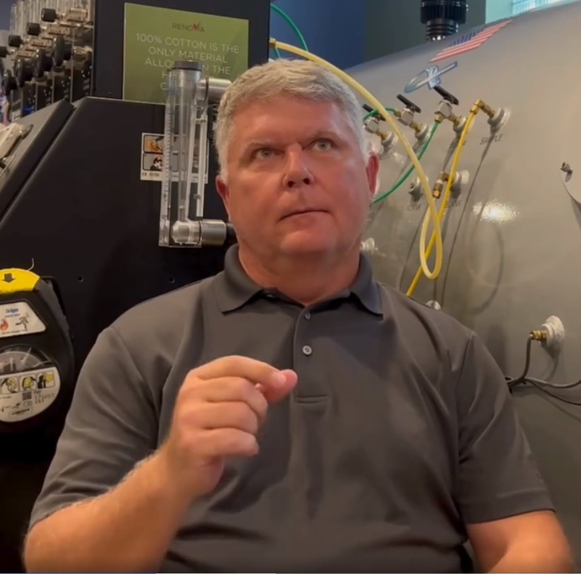
To significantly increase the delivery of oxygen delivery to the tissues requires the use of hyperbaric conditions, that is, pressures greater than normal sea level atmospheric pressure. When tissue is damaged the blood supply within the tissue is also damaged and too little oxygen may be available for recovery to take place. Hyperbaric medicine is not taught in most medical schools and is often dismissed by doctors as “alternative” medicine, but it is drugs that are alternative. Some raise fears about toxicity but in practice this is not a problem. More is known about oxygen and its dosage than any pharmaceutical. There is no more important intervention than to give sufficient oxygen to correct a tissue deficiency but, unfortunately, oxygen is only given in hospital to restore normal levels in the blood. The increased pressure has no effect on the body, although the pressure in the middle ear and sinuses in adults has to be equalized.
More oxygen may help many children with cerebral palsy, but it is not a cure. There are some obvious questions to be answered:
WHEN DOES THE DAMAGE OCCUR?
Ultrasonic scanning of the brain has shown that in most children the events which cause the development of cerebral palsy (CP) occur at the time of birth 1, although it may be many months before spasticity develops.2 Where does the damage occur? The areas affected in CP are in the middle of the hemispheres of the brain and one side or both sides may involved. These critical areas, called the internal capsules, are where the fibres from the controlling nerve cells in the grey matter of the brain pass down on their way to the spinal cord. In the spinal cord they interconnect with the nerve cells whose fibres activate the muscles of the legs and arms.
WHY DOES THE DAMAGE OCCUR?
Unfortunately, the internal capsules have a poor blood supply, shown by the frequent occurrence of damage to these areas in younger patients with multiple sclerosis and in strokes in the elderly by Magnetic Resonance Imaging (MRI). When any event causes lack of oxygen the blood vessels leak, the tissues become swollen and there may even be leakage of blood. The increased water content, termed oedema, reduces the transport of oxygen. This applies to any tissue, but especially to the brain where a sufficient quantity of oxygen is vital both to the function and, in children, its development. What causes paralysis and spasticity to develop? When the controlling nerve cells in the brain
are disconnected from the spinal cord, the signals to the arms and legs cannot pass and the ability to move is lost. Eventually, because the nerve cells in the spinal cord are separated from the control of the brain, they send an excess of signals to the muscles, causing the uncontrolled contractions known as spasticity. The areas carrying the nerve fibres to the legs are the closest to the ventricles of the brain where the blood supply is poorest3 so the legs are the most commonly affected. The is called diplegia, to indicate that the problem is in the brain and distinguish it from paraplegia where the damage is in the spinal cord.
WHY IS SPASTICITY DELAYED?
This is a crucial question that is, at present, not adequately explained or even raised. Children who develop spasticity often appear to develop normally for several months and then lose function gradually. Because in many children there is voluntary movement for a time after birth, the connections must still be intact. Why then are they lost allowing spasticity to develop? The answer almost certainly is due to the failure of the coverings of the nerve fibres, known as myelin sheaths, to develop. This evidence has come from MRI.2 Myelin sheaths envelop the nerve fibres like a Swiss roll in order to increase the speed of impulse transmission. Myelination normally begins about a month before birth and progresses to completion by the age of two. If there is tissue swelling in the mid-brain the delicate cells that form myelin die and the nerve fibres, left exposed, slowly deteriorate with the ultimate development of spasticity.
WHAT MAY BE POSSIBLE?
Loss of function in the brain can be either due to tissue swelling, which is reversible, or tissue destruction, which is not. The recoverable areas can now be identified by a technique called SPECT imaging. The initials stand for Single Photon Emission Computed Tomography. It can demonstrate blood flow which is linked to metabolism of the brain which is, of course, directly related to oxygen availability. By giving oxygen at the high dosages possible under hyperbaric conditions, areas which are not ”dead but sleeping” can be identified. This phenomenon has been discussed for many years in stroke patients and authorities have even stated that the critical parameter is not blood flow it is oxygen delivery.4 Under normal circumstances, blood flow and oxygen delivery are inextricably coupled, but the use of hyperbaric conditions can change this situation. Tissue oedema and swelling may persist in, for example, joints, for many years and SPECT imaging has now revealed that this is true in the brain.5 Suggesting that more oxygen, that is additional oxygen supplied under hyperbaric conditions may be of value generates further questions:
WHAT DOES HYPERBARIC MEAN?
It means a pressure greater than normal sea-level atmospheric pressure. Atmospheric pressure at sea-level varies with the weather and on a high pressure day more oxygen is available to the body. Aches and pains may be worse on a low pressure day because of the reduction of oxygen pressure. A hyperbaric chamber allows much more oxygen to be dissolved in the blood. An indication of the power of this technique is that at twice atmospheric pressure breathing pure oxygen the work of the heart is reduced by 20%. So much can be dissolved in the plasma that life is possible for a short time without red blood cells. The research behind the development of hyperbaric oxygen therapy has been undertaken by doctors involved in aviation, space exploration and diving. This critical information is not yet taught in our Medical Schools, despite many thousands of published articles including controlled studies in many conditions.
HOW CAN CEREBRAL PALSY CHILDREN BE HELPED?
Clearly the appropriate time to use of oxygen is at the start of a disease process, not after a delay of months or years. Nevertheless, a course of oxygen therapy sessions at increased pressure has been shown to resolve tissue swelling after the lapse of years. It works by constricting blood vessels and interrupting the vicious cycle where oxygen lack leads to tissue swelling, which then leads to further oxygen deficiency. Although formal studies have yet to be undertaken in children with cerebral palsy there is every reason to believe that exactly the same effect that is seen in stroke patients can occur. Also in children the brain is still developing and therefore the prospects for improvement are very much greater than in adults. Recovery of brain damage in children resulting from cardiac surgery has been documented using X-ray scanning.6
WILL OXYGEN THERAPY CURE CEREBRAL PALSY?
Hyperbaric oxygen therapy is not a miracle cure for children with cerebral palsy, it is simply a way of ensuring the most complete recovery possible. It should be used with exercise programmes, because lack of use in muscles and joints leads to changes that can only be reversed by exercise.
WHY ARE THERE NO FORMAL STUDIES?
Formal studies are now underway in the USA and Canada and the results of the pilot study in McGill University are now ready for publication. There is a first time for everything. Unfortunately most of the medical research in the UK is funded by the drug industry and the
costs involved are enormous. As the use of oxygen cannot be patented, there is no way that the cost of trials could be recouped and no finance is available for the promotion of the therapy. Because of the great advances made in the use of drugs a climate has been created in which doctors are conditioned to expect a drug-based solution to every disease. Oxygen has been available in Medicine for over a hundred years so it is difficult to accept that it is not being used properly, but over 500 chambers are now operating in the USA and Japan, 1500 in Russia and a similar number in China. As is so often the case much of the original research was undertaken and published in the UK. In many diseases the cost of investigations is often a great deal more than the cost of providing hyperbaric oxygen therapy. MRI and SPECT imaging may allow the benefit to be demonstrated, but they are not in any way therapeutic. There is no better assessor of a child suffering from cerebral palsy than a parent or carer involved in day-to-day hands on care.
ARE THERE DANGERS ?
The only risk with hyperbaric conditions properly supervised is to the ear drum, just as when aircraft – which are hyperbaric chambers – descend. There are limits to oxygen delivery, for example, the very high pressures used in diving can cause convulsions, but the Chinese have shown that epilepsy is actually treated by hyperbaric oxygen therapy at lower pressures. There is no evidence of either eye or lung toxicity at 1.5-1.75 atm abs.
References
- Pape KE, Wiggleworth JS. Haemorrhage, ischaemia and the perinatal brain. Clinics in developmental medicine. Nos. 69/70 William Heinemann Medical Books, London, 1979.
- Dubowitz LMS, Bydder GM, Mushin J. Developmental sequence of periventricular leukomalacia. Arch Dis Child 1985;60:349-55.
- Takashima S, Tanaka K. Development of cerebrovascular architecture and its relationship to periventricular leukomalacia. Arch Neurol 1978;35:11-16.
- Astrup J, Siesjo BK, Symon L. Thresholds in cerebral ischemia; the ischemic penumbra. Stroke 1981;12:723-25.
- Neubauer RA, Gottlieb SF, Kagan RL. Enhancing idling neurones. Lancet 1990;336:542.
- Muraoka R, Yokota M, Aoshima M, et al. Subclinical changes in brain morphology following cardiac operations as reflected by computed tomographic scans of the brain. J Thorac Cardiovasc Surg 1981;81:364-69.


